File:Acute portal vein thrombosis (Radiopaedia 73198-83925 Axial Heavily T2 thin cuts 6).jpg
Jump to navigation
Jump to search
No higher resolution available.
Acute_portal_vein_thrombosis_(Radiopaedia_73198-83925_Axial_Heavily_T2_thin_cuts_6).jpg (635 × 476 pixels, file size: 136 KB, MIME type: image/jpeg)
Summary:
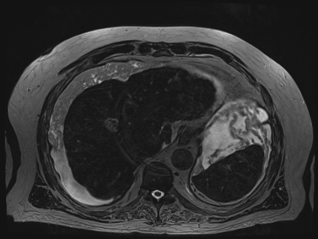
| Description |
|
| Date | Published: 22nd Jan 2020 |
| Source | https://radiopaedia.org/cases/acute-portal-vein-thrombosis |
| Author | Mostafa El-Feky |
| Permission (Permission-reusing-text) |
http://creativecommons.org/licenses/by-nc-sa/3.0/ |
Licensing:
Attribution-NonCommercial-ShareAlike 3.0 Unported (CC BY-NC-SA 3.0)
File history
Click on a date/time to view the file as it appeared at that time.
| Date/Time | Thumbnail | Dimensions | User | Comment | |
|---|---|---|---|---|---|
| current | 07:02, 18 April 2021 | | 635 × 476 (136 KB) | Fæ (talk | contribs) | Radiopaedia project rID:73198 (batch #1043-277 G6) |
You cannot overwrite this file.
File usage
The following page uses this file:
.jpg&oldid=159319)