File:Acute pulmonary edema, mitral regurgitation and mucopolysaccharidosis (Radiopaedia 56239).jpg
Jump to navigation
Jump to search

Size of this preview: 468 × 599 pixels. Other resolutions: 187 × 240 pixels | 375 × 480 pixels | 600 × 768 pixels | 800 × 1,024 pixels | 1,534 × 1,964 pixels.
Original file (1,534 × 1,964 pixels, file size: 843 KB, MIME type: image/jpeg)
Summary:
- Radiopaedia case ID: 56239
- Image ID: 33385982
- Modality: X-ray
- System: Musculoskeletal
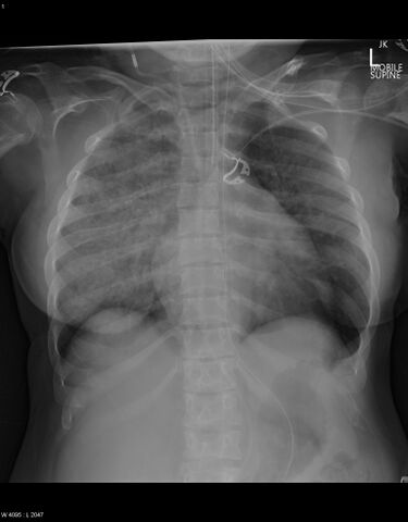
- Findings: The ETT and NGT are well-positioned. Tubing through the right side of the neck and over the midline of the chest likely represents VP shunt tubing which is intact. The heart is enlarged even allowing for projection, and there is abnormal cardiac contour, in keeping with history of mitral valve disease. There is diffuse air space opacification, more so on the right side, with perihilar infiltrates. No pleural effusion or evidence of pneumothorax. Features are in keeping with asymmetric acute pulmonary edema. Posterior ribs are abnormal, in keeping with history of MPS.
- Published: 20th Oct 2017
- Source: https://radiopaedia.org/cases/acute-pulmonary-oedema-mitral-regurgitation-and-mucopolysaccharidosis
- Author: Craig Hacking
- Permission: http://creativecommons.org/licenses/by-nc-sa/3.0/
Licensing:
Attribution-NonCommercial-ShareAlike 3.0 Unported (CC BY-NC-SA 3.0)
File history
Click on a date/time to view the file as it appeared at that time.
| Date/Time | Thumbnail | Dimensions | User | Comment | |
|---|---|---|---|---|---|
| current | 16:16, 18 March 2021 | | 1,534 × 1,964 (843 KB) | Fæ (talk | contribs) | Radiopaedia project rID:56239 (batch #1048) |
You cannot overwrite this file.
File usage
There are no pages that use this file.
.jpg&oldid=8859946)