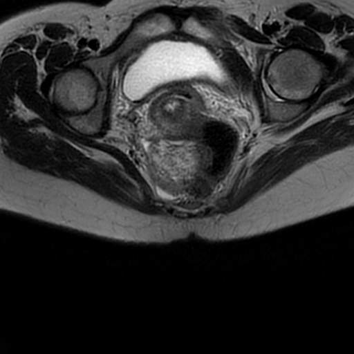
File:Acute sacroiliitis (Radiopaedia 67272-76633 Axial T2 19).jpg
Jump to navigation
Jump to search
Acute_sacroiliitis_(Radiopaedia_67272-76633_Axial_T2_19).jpg (512 × 512 pixels, file size: 22 KB, MIME type: image/jpeg)
Summary:
| Description |
|
| Date | Published: 26th Mar 2019 |
| Source | https://radiopaedia.org/cases/acute-sacroiliitis-1 |
| Author | Heba Abdelmonem |
| Permission (Permission-reusing-text) |
http://creativecommons.org/licenses/by-nc-sa/3.0/ |
Licensing:
Attribution-NonCommercial-ShareAlike 3.0 Unported (CC BY-NC-SA 3.0)
File history
Click on a date/time to view the file as it appeared at that time.
| Date/Time | Thumbnail | Dimensions | User | Comment | |
|---|---|---|---|---|---|
| current | 07:17, 19 April 2021 |  | 512 × 512 (22 KB) | Fæ (talk | contribs) | Radiopaedia project rID:67272 (batch #1093-43 B19) |
You cannot overwrite this file.
File usage
There are no pages that use this file.
