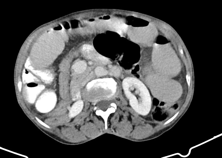
File:Acute small bowel (ileal) volvulus (Radiopaedia 71740-82139 Axial C+ portal venous phase 90).jpg
Jump to navigation
Jump to search
Acute_small_bowel_(ileal)_volvulus_(Radiopaedia_71740-82139_Axial_C+_portal_venous_phase_90).jpg (718 × 512 pixels, file size: 52 KB, MIME type: image/jpeg)
Summary:
| Description |
|
| Date | 27 Oct 2019 |
| Source | Acute small bowel (ileal) volvulus |
| Author | Michael P Hartung |
| Permission (Permission-reusing-text) |
http://creativecommons.org/licenses/by-nc-sa/3.0/ |
Licensing:
Attribution-NonCommercial-ShareAlike 3.0 Unported (CC BY-NC-SA 3.0)
| This file is ineligible for copyright and therefore in the public domain, because it is a technical image created as part of a standard medical diagnostic procedure. No creative element rising above the threshold of originality was involved in its production.
|
File history
Click on a date/time to view the file as it appeared at that time.
| Date/Time | Thumbnail | Dimensions | User | Comment | |
|---|---|---|---|---|---|
| current | 15:28, 19 April 2021 |  | 718 × 512 (52 KB) | Fæ (talk | contribs) | Radiopaedia project rID:71740 (batch #1108-90 A90) |
You cannot overwrite this file.
File usage
The following page uses this file:

