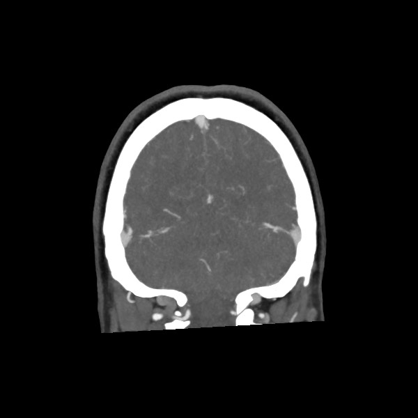
File:Acute subarachnoid hemorrhage and accessory anterior cerebral artery (Radiopaedia 69231-79009 Coronal C+ arterial phase 65).jpg
Jump to navigation
Jump to search
No higher resolution available.
Acute_subarachnoid_hemorrhage_and_accessory_anterior_cerebral_artery_(Radiopaedia_69231-79009_Coronal_C+_arterial_phase_65).jpg (600 × 600 pixels, file size: 27 KB, MIME type: image/jpeg)
Summary:
| Description |
|
| Date | Published: 3rd Jul 2019 |
| Source | https://radiopaedia.org/cases/acute-subarachnoid-haemorrhage-and-accessory-anterior-cerebral-artery |
| Author | Madeleine Scicchitano |
| Permission (Permission-reusing-text) |
http://creativecommons.org/licenses/by-nc-sa/3.0/ |
Licensing:
Attribution-NonCommercial-ShareAlike 3.0 Unported (CC BY-NC-SA 3.0)
File history
Click on a date/time to view the file as it appeared at that time.
| Date/Time | Thumbnail | Dimensions | User | Comment | |
|---|---|---|---|---|---|
| current | 18:01, 19 April 2021 | | 600 × 600 (27 KB) | Fæ (talk | contribs) | Radiopaedia project rID:69231 (batch #1112-195 B65) |
You cannot overwrite this file.
File usage
The following page uses this file:
.jpg&oldid=169441)