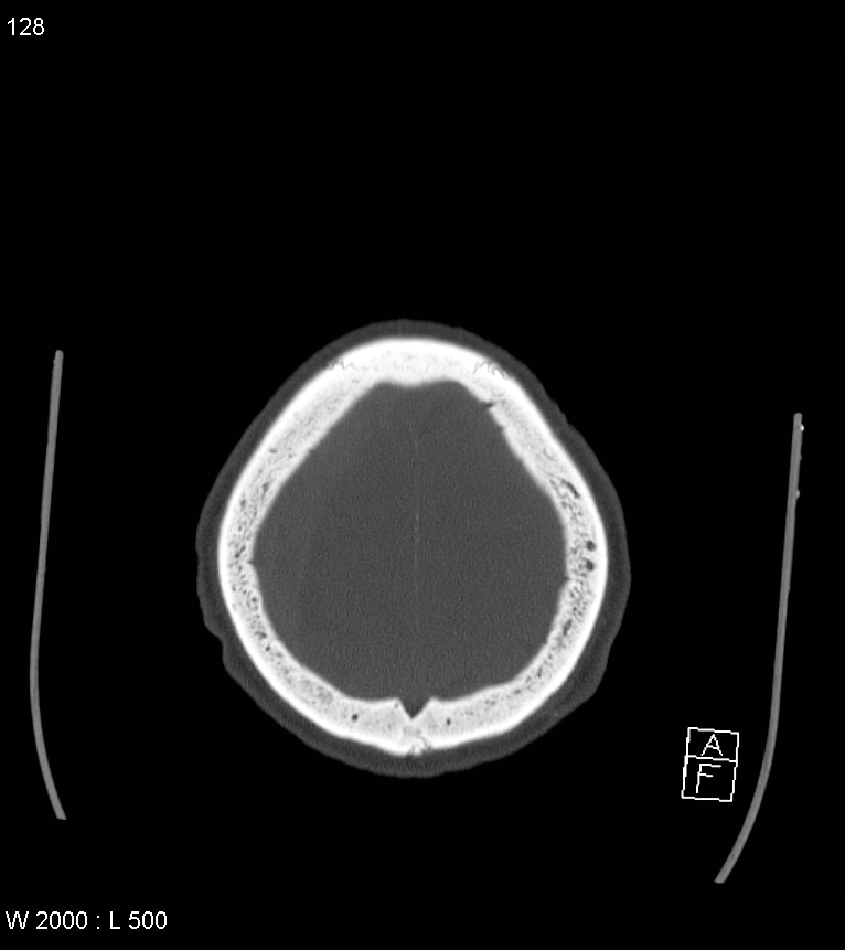
File:Acute subdural hematoma with myelofibrosis (Radiopaedia 52582-58494 Axial bone window 64).jpg
Jump to navigation
Jump to search
Size of this preview: 533 × 599 pixels. Other resolutions: 213 × 240 pixels | 427 × 480 pixels | 766 × 861 pixels.
Original file (766 × 861 pixels, file size: 97 KB, MIME type: image/jpeg)
Summary:
| Description |
|
| Date | Published: 1st Jun 2017 |
| Source | https://radiopaedia.org/cases/acute-subdural-haematoma-with-myelofibrosis |
| Author | Craig Hacking |
| Permission (Permission-reusing-text) |
http://creativecommons.org/licenses/by-nc-sa/3.0/ |
Licensing:
Attribution-NonCommercial-ShareAlike 3.0 Unported (CC BY-NC-SA 3.0)
File history
Click on a date/time to view the file as it appeared at that time.
| Date/Time | Thumbnail | Dimensions | User | Comment | |
|---|---|---|---|---|---|
| current | 20:20, 19 April 2021 | | 766 × 861 (97 KB) | Fæ (talk | contribs) | Radiopaedia project rID:52582 (batch #1116-215 D64) |
You cannot overwrite this file.
File usage
The following page uses this file:
.jpg&oldid=170251)