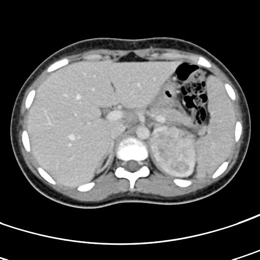
File:Acute suppurative pyelonephritis (Radiopaedia 18306-18147 Axial renal parenchymal phase 41).jpg
Jump to navigation
Jump to search
Acute_suppurative_pyelonephritis_(Radiopaedia_18306-18147_Axial_renal_parenchymal_phase_41).jpg (512 × 512 pixels, file size: 28 KB, MIME type: image/jpeg)
Summary:
| Description |
|
| Date | Published: 28th Jun 2012 |
| Source | https://radiopaedia.org/cases/acute-suppurative-pyelonephritis |
| Author | Hani Makky Al Salam |
| Permission (Permission-reusing-text) |
http://creativecommons.org/licenses/by-nc-sa/3.0/ |
Licensing:
Attribution-NonCommercial-ShareAlike 3.0 Unported (CC BY-NC-SA 3.0)
File history
Click on a date/time to view the file as it appeared at that time.
| Date/Time | Thumbnail | Dimensions | User | Comment | |
|---|---|---|---|---|---|
| current | 01:55, 20 April 2021 |  | 512 × 512 (28 KB) | Fæ (talk | contribs) | Radiopaedia project rID:18306 (batch #1134-41 A41) |
You cannot overwrite this file.
File usage
The following page uses this file:
