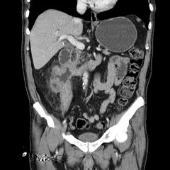
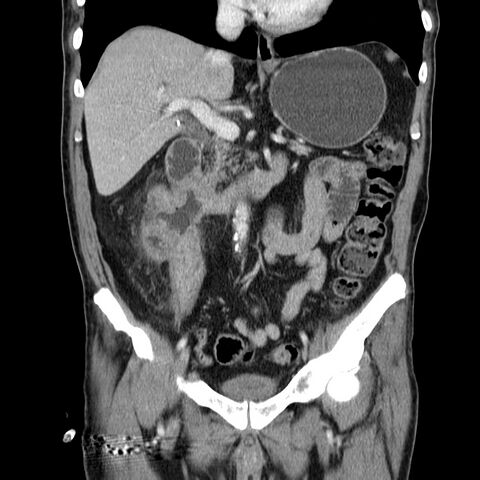
File:Adenocarcinoma of colon with entero-colic fistula (Radiopaedia 22832-22852 Coronal C+ portal venous phase 26).jpg
Jump to navigation
Jump to search

Size of this preview: 600 × 600 pixels. Other resolutions: 240 × 240 pixels | 480 × 480 pixels | 840 × 840 pixels.
Original file (840 × 840 pixels, file size: 181 KB, MIME type: image/jpeg)
Summary:
| Description |
|
| Date | Published: 27th Apr 2013 |
| Source | https://radiopaedia.org/cases/adenocarcinoma-of-colon-with-entero-colic-fistula |
| Author | David Cuete |
| Permission (Permission-reusing-text) |
http://creativecommons.org/licenses/by-nc-sa/3.0/ |
Licensing:
Attribution-NonCommercial-ShareAlike 3.0 Unported (CC BY-NC-SA 3.0)
File history
Click on a date/time to view the file as it appeared at that time.
| Date/Time | Thumbnail | Dimensions | User | Comment | |
|---|---|---|---|---|---|
| current | 23:34, 21 April 2021 | | 840 × 840 (181 KB) | Fæ (talk | contribs) | Radiopaedia project rID:22832 (batch #1178-102 B26) |
You cannot overwrite this file.
File usage
The following page uses this file:
.jpg&oldid=186970)