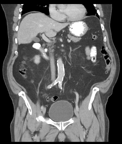
File:Adenocarcioma of rectum- T1 lesion (Radiopaedia 36921-38547 Coronal C+ portal venous phase 30).png

From NC Commons
Jump to navigation Jump to search

Original file(512 × 603 pixels, file size: 280 KB, MIME type: image/png)

Summary:

Description
  • Radiopaedia case ID: 36921
  • Image ID: 12734463
  • Image stack position: 30/63
  • Plane projection: Coronal
  • Aux modality: C+ portal venous phase
  • Study description: Staging CT
  • Study findings: CT Chest, Abdomen and Pelvis. Dynamically enhanced axial images have been obtained through the chest, abdomen and pelvis following intravenous and oral contrast. Within the chest, a 5mm nodule abuts the oblique fissure in the anterior aspect of the left lower lobe. Minimal atelectasis is seen in base of the left lingula lobe. No pleural or pericardial effusion. No mediastinal, hilar, axillary or retrocrural lymphadenopathy. Within the abdomen and pelvis, the liver is of mildly decreased attenuation suggesting fatty infiltration. No focal liver lesion is seen. The spleen, pancreas and adrenal glands have normal appearance. Small bilateral low-attenuation renal lesions have CT features consistent with cysts. The largest lies in the mid pole of the right kidney measuring approximately 15 mm, with exophytic 13 mm cyst in the inferior pole of the left kidney. No hydronephrosis. Multiple small and borderline in size and left para-aortic lymph nodes are demonstrated. None of which however has significantly enlarged on size criteria. There is no mesenteric or inguinal lymphadenopathy. No free fluid or focal collection is seen within the abdomen or pelvis. Colonic diverticular disease is demonstrated. No evidence of bowel obstruction. The known rectal lesion is not clearly identified. The surrounding perirectal fat is preserved. No suspicious bony lesion. Conclusion A 5 mm left lower lobe pulmonary nodule is non-specific. Correlation, with an early followup study in 3 months time or comparison with any prior imaging is suggested. There is no definite evidence of metastatic disease within the chest, abdomen or pelvis.
  • Modality: CT
  • System: Gastrointestinal
  • Findings: Malignant appearing rectal polypoid lesion
Date Published: 19th May 2015
Source https://radiopaedia.org/cases/adenocarcioma-of-rectum-t1-lesion
Author Jan Frank Gerstenmaier
Permission
(Permission-reusing-text)
http://creativecommons.org/licenses/by-nc-sa/3.0/

Licensing:

Attribution-NonCommercial-ShareAlike 3.0 Unported (CC BY-NC-SA 3.0)

File history

Click on a date/time to view the file as it appeared at that time.

Date/TimeThumbnailDimensionsUserComment
current02:10, 23 April 2021Thumbnail for version as of 02:10, 23 April 2021512 × 603 (280 KB) (talk | contribs)Radiopaedia project rID:36921 (batch #1196-131 B30)

Metadata