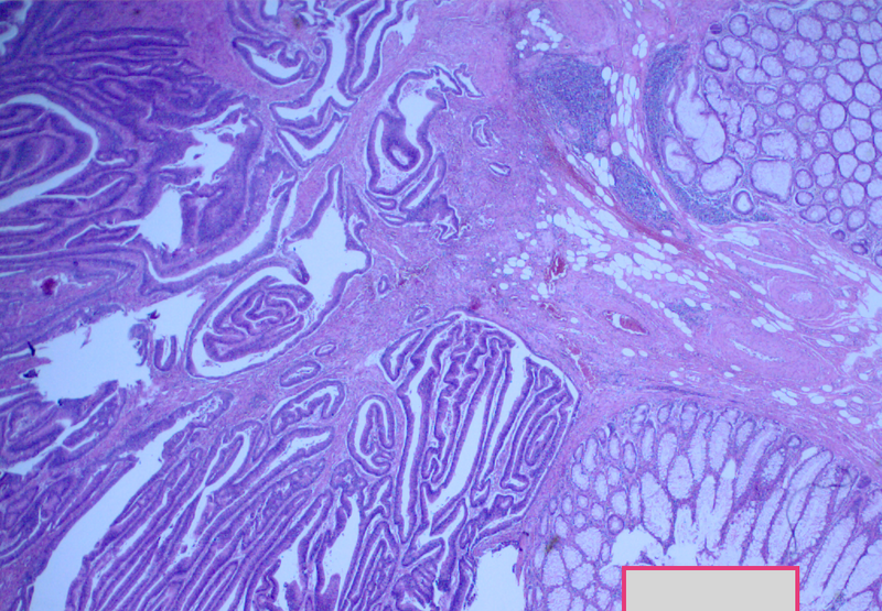
File:Adenocarcioma of rectum- T1 lesion (Radiopaedia 36921-38552 A 1).PNG

From NC Commons
Jump to navigation Jump to search

Original file(1,017 × 705 pixels, file size: 1.64 MB, MIME type: image/png)

Summary:

Description
  • Radiopaedia case ID: 36921
  • Image ID: 12735124
  • Study findings: CLINICAL NOTES: 1. Rectal cancer Ultra low anterior resection 2. Distal Donut MACROSCOPIC DESCRIPTION: 1. "Rectum": A 275mm length anterior resection, diameter 20-30mm. The anterior peritoneal reflection is 30mm from distal margin. The posterior perirectal fat is non-peritonealised from 170mm from distal margin. There is up to 100mm mesocolon, up to 29mm posterior perirectal fat, up to 20mm anterior perirectal fat. Within the rectum, at the level of the anterior peritoneal reflection, (22mm from distal margin and 212mm from proximal margin), there is a pale brown smooth discoid tumor, 20x13x9mm. The tumor is causing a puckering of the underlying mucosa but appears to be limited to mucosa and submucosa, with intact muscularis propria. Tumor submitted in full. The tumor is on posterior wall of rectum, where there is up to 19mm of posterior perirectal fat (tumor is 30mm from posterior non-peritonealised surface). The distal 120mm of rectum shows diffuse polypoid lesions, 2-5mm maximum dimension (approximately 45 in total). The polyps are present at distal margin, and 155mm from proximal margin. The sigmoid mucosa contains a single 4mm diameter polyp,and is otherwise shiny and unremarkable. Representative sections of largest polyps submitted. Inking notation: proximal margin black, distal margin black, anterior circumferential margin blue, posterior circumferential margin green. BLOCK DESIGNATION: 1A - proximal margin. 1B - distal margin. 1C-1E - rectal tumor (submitted in full). 1F - largest rectal polyp bisected. 1G - four rectal polyps. 1H - seven whole nodes. 1I - six whole nodes. P9. 2. "Distal donut": A mucosal donut, 22x10mm, length 9mm. Staples removed. Longitudinal sections of mucosa submitted. P1. (CH) MICROSCOPIC DESCRIPTION: 1. The sections show invasive moderately differentiated adenocarcinoma arising in a tubulovillous adenoma with high grade dysplasia involving rectal mucosa. Tumor extends into the submucosa but does not appear to involve muscularis propria. No evidence of vascular, lymphatic or perineural invasion is seen. Of the five other polypoid lesions sampled, one is a sessile serrated adenoma and the remaining four are hyperplastic polyps. No evidence of metastatic tumor is seen in any of 10 lymph nodes. All resection margins are clear of tumor. 2. The section shows rectal mucosa and wall. A small hyperplastic polyp is noted within rectal mucosa. No evidence of invasive tumor is seen. DIAGNOSIS: 1. Rectum: Site - rectum. Type - moderately differentiated adenocarcinoma arising in tubulovillous adenoma with high grade dysplasia. Size - 20mm in maximum dimension. Local invasion - tumor extends through muscularis mucosae into submucosa; no evidence of involvement of muscularis propria seen. Lymphovascular invasion - absent. Perineural invasion - absent. Proximal, distal, radial - all clear of tumor. Lymph nodes - no evidence of metastatic tumor in any of 10 lymph nodes. Additional pathology - five sampled polypoid lesions - 1x sessile serrated adenoma; 4x hyperplastic polyp. AJCC (7th edition) Stage 1 (T1 N0 MX). *** Identification and sectioning of further lymph nodes pending Carnoy's treatment of perirectal fat. 2. Distal donut: Rectal mucosa with small hyperplastic polyp; no evidence of invasive tumor seen.
  • Modality: Pathology
  • System: Gastrointestinal
  • Findings: Malignant appearing rectal polypoid lesion
Date Published: 19th May 2015
Source https://radiopaedia.org/cases/adenocarcioma-of-rectum-t1-lesion
Author Jan Frank Gerstenmaier
Permission
(Permission-reusing-text)
http://creativecommons.org/licenses/by-nc-sa/3.0/

Licensing:

Attribution-NonCommercial-ShareAlike 3.0 Unported (CC BY-NC-SA 3.0)

File history

Click on a date/time to view the file as it appeared at that time.

Date/TimeThumbnailDimensionsUserComment
current03:37, 23 April 2021Thumbnail for version as of 03:37, 23 April 20211,017 × 705 (1.64 MB) (talk | contribs)Radiopaedia project rID:36921 (batch #1196-1 A1)

There are no pages that use this file.