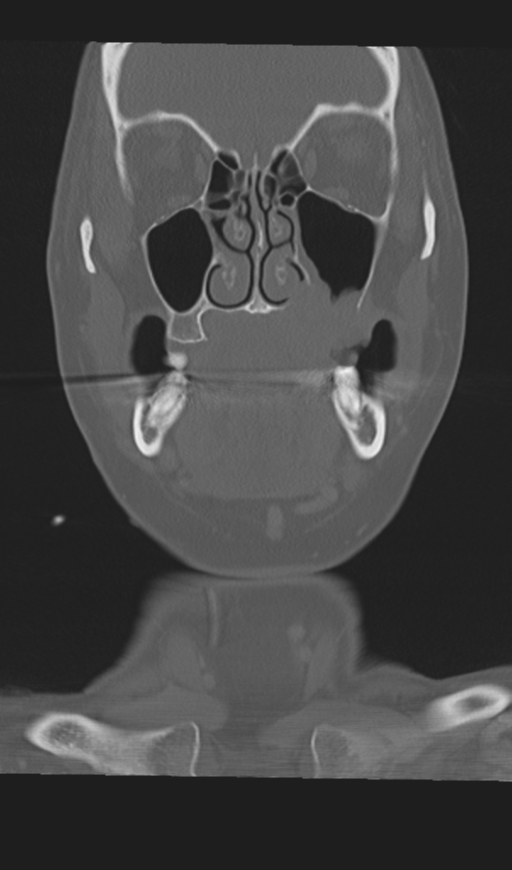
File:Adenoid cystic tumor of palate (Radiopaedia 46980-51518 Coronal bone window 23).png
Jump to navigation
Jump to search
Size of this preview: 353 × 600 pixels. Other resolutions: 141 × 240 pixels | 512 × 870 pixels.
Original file (512 × 870 pixels, file size: 131 KB, MIME type: image/png)
Summary:
| Description |
|
| Date | Published: 1st Aug 2016 |
| Source | https://radiopaedia.org/cases/adenoid-cystic-tumour-of-palate |
| Author | Melbourne Uni Radiology Masters |
| Permission (Permission-reusing-text) |
http://creativecommons.org/licenses/by-nc-sa/3.0/ |
Licensing:
Attribution-NonCommercial-ShareAlike 3.0 Unported (CC BY-NC-SA 3.0)
File history
Click on a date/time to view the file as it appeared at that time.
| Date/Time | Thumbnail | Dimensions | User | Comment | |
|---|---|---|---|---|---|
| current | 23:00, 23 April 2021 | | 512 × 870 (131 KB) | Fæ (talk | contribs) | Radiopaedia project rID:46980 (batch #1218-236 D23) |
You cannot overwrite this file.
File usage
The following page uses this file:
.png&oldid=201907)