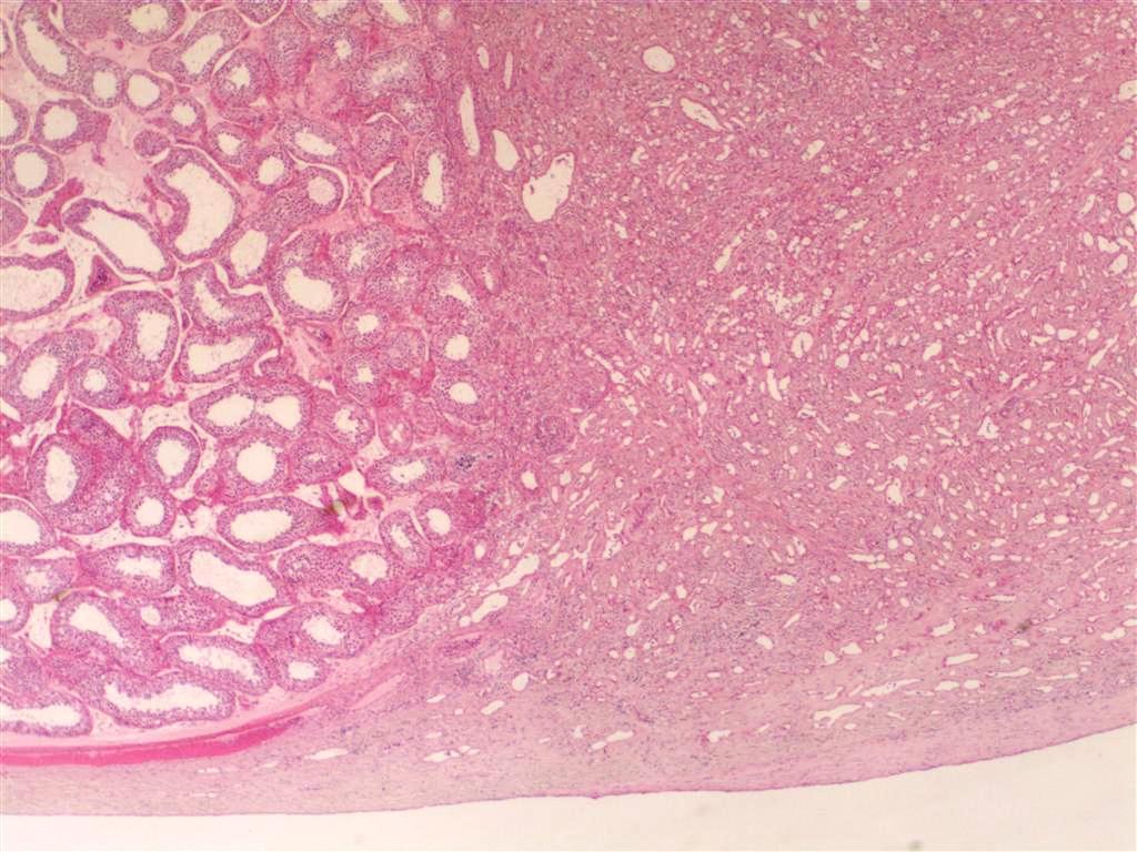
File:Adenomatoid tumor of the scrotum (pathology) (Radiopaedia 16175-15853 H&E 1).jpg
Jump to navigation
Jump to search
Size of this preview: 800 × 600 pixels. Other resolutions: 320 × 240 pixels | 640 × 480 pixels | 1,024 × 768 pixels.
Original file (1,024 × 768 pixels, file size: 141 KB, MIME type: image/jpeg)
Summary:
| Description |
|
| Date | Published: 27th Dec 2011 |
| Source | https://radiopaedia.org/cases/adenomatoid-tumour-of-the-scrotum-pathology |
| Author | Andrew Ryan |
| Permission (Permission-reusing-text) |
http://creativecommons.org/licenses/by-nc-sa/3.0/ |
Licensing:
Attribution-NonCommercial-ShareAlike 3.0 Unported (CC BY-NC-SA 3.0)
File history
Click on a date/time to view the file as it appeared at that time.
| Date/Time | Thumbnail | Dimensions | User | Comment | |
|---|---|---|---|---|---|
| current | 04:27, 24 April 2021 | | 1,024 × 768 (141 KB) | Fæ (talk | contribs) | Radiopaedia project rID:16175 (batch #1229-2 B1) |
You cannot overwrite this file.
File usage
There are no pages that use this file.
_(Radiopaedia_16175-15853_H%26E_1).jpg&oldid=203725)