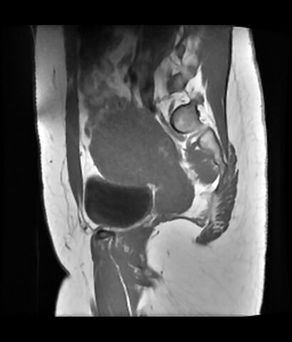
File:Adenomyosis (Radiopaedia 65071-74064 Sagittal T1 5).jpg
Jump to navigation
Jump to search

Size of this preview: 512 × 600 pixels. Other resolutions: 205 × 240 pixels | 410 × 480 pixels | 655 × 768 pixels | 874 × 1,024 pixels | 1,630 × 1,910 pixels.
Original file (1,630 × 1,910 pixels, file size: 183 KB, MIME type: image/jpeg)
Summary:
| Description |
|
| Date | Published: 21st Dec 2018 |
| Source | https://radiopaedia.org/cases/adenomyosis-12 |
| Author | Fakhry Mahmoud Ebouda |
| Permission (Permission-reusing-text) |
http://creativecommons.org/licenses/by-nc-sa/3.0/ |
Licensing:
Attribution-NonCommercial-ShareAlike 3.0 Unported (CC BY-NC-SA 3.0)
File history
Click on a date/time to view the file as it appeared at that time.
| Date/Time | Thumbnail | Dimensions | User | Comment | |
|---|---|---|---|---|---|
| current | 06:44, 24 April 2021 | | 1,630 × 1,910 (183 KB) | Fæ (talk | contribs) | Radiopaedia project rID:65071 (batch #1253-5 A5) |
You cannot overwrite this file.
File usage
There are no pages that use this file.
.jpg&oldid=204426)