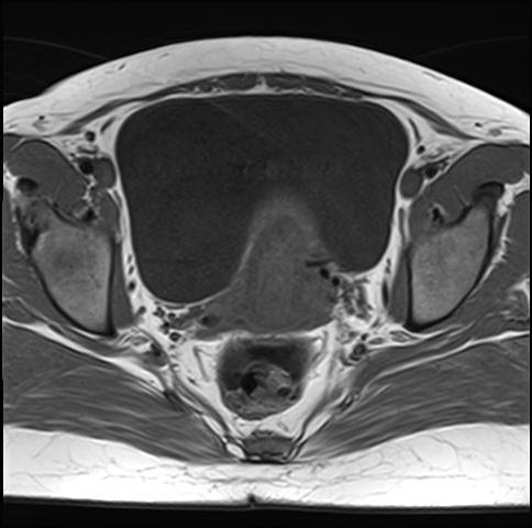
File:Adenomyosis - ovarian endometriomas (Radiopaedia 67031-76350 Axial T1 18).jpg
Jump to navigation
Jump to search
Adenomyosis_-_ovarian_endometriomas_(Radiopaedia_67031-76350_Axial_T1_18).jpg (484 × 480 pixels, file size: 102 KB, MIME type: image/jpeg)
Summary:
| Description |
|
| Date | Published: 17th Mar 2019 |
| Source | https://radiopaedia.org/cases/adenomyosis-ovarian-endometriomas |
| Author | Dr Ammar Haouimi |
| Permission (Permission-reusing-text) |
http://creativecommons.org/licenses/by-nc-sa/3.0/ |
Licensing:
Attribution-NonCommercial-ShareAlike 3.0 Unported (CC BY-NC-SA 3.0)
File history
Click on a date/time to view the file as it appeared at that time.
| Date/Time | Thumbnail | Dimensions | User | Comment | |
|---|---|---|---|---|---|
| current | 11:36, 24 April 2021 |  | 484 × 480 (102 KB) | Fæ (talk | contribs) | Radiopaedia project rID:67031 (batch #1265-18 A18) |
You cannot overwrite this file.
File usage
There are no pages that use this file.
