File:Adenomyosis of the uterus (gross pathology) (Radiopaedia 10172).jpeg
Jump to navigation
Jump to search

Size of this preview: 600 × 600 pixels. Other resolutions: 240 × 240 pixels | 480 × 480 pixels | 768 × 768 pixels | 1,024 × 1,024 pixels.
Original file (1,024 × 1,024 pixels, file size: 479 KB, MIME type: image/jpeg)
Summary:
- Radiopaedia case ID: 10172
- Image ID: 505086
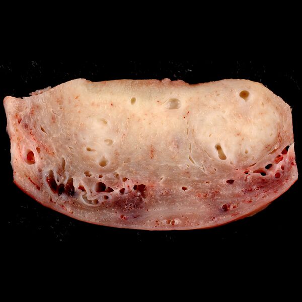
- Study findings: Cross section through the wall of a hysterectomy specimen of a 30-year-old woman who reported chronic pelvic pain and abnormal uterine bleeding. The endometrial surface is at the top of the image, and the serosa is at the bottom. The two pale areas represent regions of adenomyosis. I think most cases of adenomyosis can be reliably diagnosed grossly by an experienced prosector examining a fixed specimen. Following formalin fixation, the soft adenomyotic areas stand out more strikingly against the firmer myometrium. Author: Ed Uthman Original file: wikimedia commons here License: This file is licensed under the Creative Commons Attribution-Share Alike 2.0 Generic license. If you believe your copyright or has been infringed please write to license@radiopaedia.org giving details of why you believe this is so.
- Modality: Pathology
- System: Gynaecology
- Findings: Cross section through the wall of a hysterectomy specimen of a 30-year-old woman who reported chronic pelvic pain and abnormal uterine bleeding. The endometrial surface is at the top of the image, and the serosa is at the bottom. The two pale areas represent regions of adenomyosis. I think most cases of adenomyosis can be reliably diagnosed grossly by an experienced prosector examining a fixed specimen. Following formalin fixation, the soft adenomyotic areas stand out more strikingly against the firmer myometrium. Author: Ed UthmanOriginal file: wikimedia commons hereLicense: This file is licensed under the Creative Commons Attribution-Share Alike 2. 0 Generic license. If you believe your copyright or has been infringed please write to license@radiopaedia. org giving details of why you believe this is so.
- Published: 18th Jul 2010
- Source: https://radiopaedia.org/cases/adenomyosis-of-the-uterus-gross-pathology-1
- Author: Ed Uthman
- Permission: http://creativecommons.org/licenses/by-nc-sa/3.0/
Licensing:
Attribution-NonCommercial-ShareAlike 3.0 Unported (CC BY-NC-SA 3.0)
| This file is ineligible for copyright and therefore in the public domain, because it is a technical image created as part of a standard medical diagnostic procedure. No creative element rising above the threshold of originality was involved in its production.
|
File history
Click on a date/time to view the file as it appeared at that time.
| Date/Time | Thumbnail | Dimensions | User | Comment | |
|---|---|---|---|---|---|
| current | 16:49, 18 March 2021 |  | 1,024 × 1,024 (479 KB) | Fæ (talk | contribs) | Radiopaedia project rID:10172 (batch #1250) |
You cannot overwrite this file.
File usage
There are no pages that use this file.
