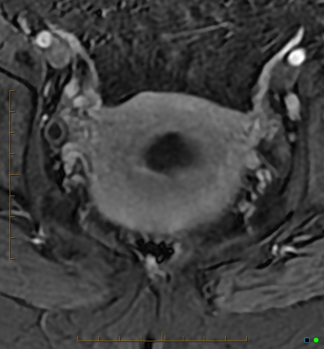
File:Adenomyosis uterus with hematometra (Radiopaedia 45779-50011 Axial T1 C+ fat sat 41).jpg
Jump to navigation
Jump to search
Size of this preview: 556 × 599 pixels. Other resolutions: 223 × 240 pixels | 634 × 683 pixels.
Original file (634 × 683 pixels, file size: 145 KB, MIME type: image/jpeg)
Summary:
| Description |
|
| Date | Published: 2nd Aug 2016 |
| Source | https://radiopaedia.org/cases/adenomyosis-uterus-with-haematometra |
| Author | Chris O'Donnell |
| Permission (Permission-reusing-text) |
http://creativecommons.org/licenses/by-nc-sa/3.0/ |
Licensing:
Attribution-NonCommercial-ShareAlike 3.0 Unported (CC BY-NC-SA 3.0)
File history
Click on a date/time to view the file as it appeared at that time.
| Date/Time | Thumbnail | Dimensions | User | Comment | |
|---|---|---|---|---|---|
| current | 13:00, 24 April 2021 | | 634 × 683 (145 KB) | Fæ (talk | contribs) | Radiopaedia project rID:45779 (batch #1267-125 D41) |
You cannot overwrite this file.
File usage
The following page uses this file:
.jpg&oldid=205496)