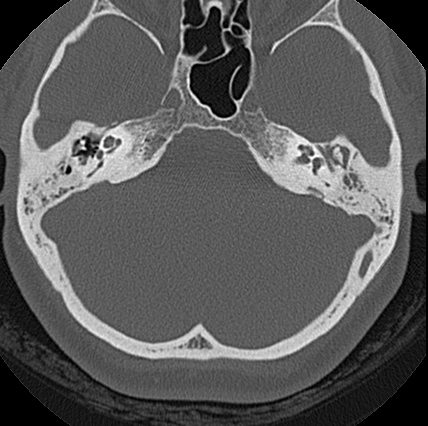
File:Adhesive chronic otitis media (Radiopaedia 14270-14148 Axial bone window 6).jpg
Jump to navigation
Jump to search
Adhesive_chronic_otitis_media_(Radiopaedia_14270-14148_Axial_bone_window_6).jpg (428 × 426 pixels, file size: 94 KB, MIME type: image/jpeg)
Summary:
| Description |
|
| Date | Published: 11th Jul 2011 |
| Source | https://radiopaedia.org/cases/adhesive-chronic-otitis-media |
| Author | Roberto Schubert |
| Permission (Permission-reusing-text) |
http://creativecommons.org/licenses/by-nc-sa/3.0/ |
Licensing:
Attribution-NonCommercial-ShareAlike 3.0 Unported (CC BY-NC-SA 3.0)
File history
Click on a date/time to view the file as it appeared at that time.
| Date/Time | Thumbnail | Dimensions | User | Comment | |
|---|---|---|---|---|---|
| current | 02:53, 25 April 2021 |  | 428 × 426 (94 KB) | Fæ (talk | contribs) | Radiopaedia project rID:14270 (batch #1295-6 A6) |
You cannot overwrite this file.
File usage
There are no pages that use this file.
