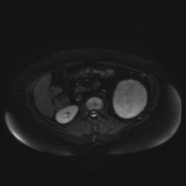
File:Adrenal cortical carcinoma (Radiopaedia 70906-81116 Axial DWI 17).jpg
Jump to navigation
Jump to search
Size of this preview: 600 × 600 pixels. Other resolutions: 240 × 240 pixels | 480 × 480 pixels | 762 × 762 pixels.
Original file (762 × 762 pixels, file size: 80 KB, MIME type: image/jpeg)
Summary:
| Description |
|
| Date | Published: 14th Sep 2019 |
| Source | https://radiopaedia.org/cases/adrenal-cortical-carcinoma-4 |
| Author | Dr Ammar Haouimi |
| Permission (Permission-reusing-text) |
http://creativecommons.org/licenses/by-nc-sa/3.0/ |
Licensing:
Attribution-NonCommercial-ShareAlike 3.0 Unported (CC BY-NC-SA 3.0)
File history
Click on a date/time to view the file as it appeared at that time.
| Date/Time | Thumbnail | Dimensions | User | Comment | |
|---|---|---|---|---|---|
| current | 07:17, 25 April 2021 | | 762 × 762 (80 KB) | Fæ (talk | contribs) | Radiopaedia project rID:70906 (batch #1320-335 J17) |
You cannot overwrite this file.
File usage
The following page uses this file:
.jpg&oldid=210469)