File:Adrenal cyst (Radiopaedia 45625-49777 Axial C+ portal venous phase 24).png

From NC Commons
Jump to navigation Jump to search

Adrenal_cyst_(Radiopaedia_45625-49777_Axial_C+_portal_venous_phase_24).png(512 × 512 pixels, file size: 190 KB, MIME type: image/png)

Summary:

Description
  • Radiopaedia case ID: 45625
  • Image ID: 22978604
  • Image stack position: 24/112
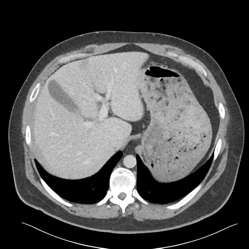
  • Plane projection: Axial
  • Aux modality: C+ portal venous phase
  • Study description: 3 years later
  • Study findings: The low-density lesion previously seen arising from the lateral limb of the right adrenal gland has increased in size now measuring up to 5.7 x 4.8 x 4.5 cm. The density has only slightly increased now measuring approximately 17 HU (Again, this is a portal venous phase study). Further assessment with adrenal washout CT is recommended to confirm suspected (benign) cystic nature of the lesion.
  • Modality: CT
  • System: Urogenital
  • Findings: Small umbilical hernia containing omental fat. There is a small amount of fat stranding in the fat just subjacent to the hernia. No bowel demonstrated within the hernia. There is a small fat-containing inguinal hernia bilaterally. Fatty infiltration of the liver. Ovoid 2.5 x 2.5 x 2.5 cm low-density lesion (8-15HU) arising from the lateral limb of the right adrenal gland is probably an adrenal adenoma (the study was only obtained in portal vein phase). The left adrenal gland is normal. The remainder study is unremarkable.
Date Published: 14th Jun 2016
Source https://radiopaedia.org/cases/adrenal-cyst
Author Bruno Di Muzio
Permission
(Permission-reusing-text)
http://creativecommons.org/licenses/by-nc-sa/3.0/

Licensing:

Attribution-NonCommercial-ShareAlike 3.0 Unported (CC BY-NC-SA 3.0)

File history

Click on a date/time to view the file as it appeared at that time.

Date/TimeThumbnailDimensionsUserComment
current16:47, 25 April 2021Thumbnail for version as of 16:47, 25 April 2021512 × 512 (190 KB) (talk | contribs)Radiopaedia project rID:45625 (batch #1327-24 A24)

Metadata