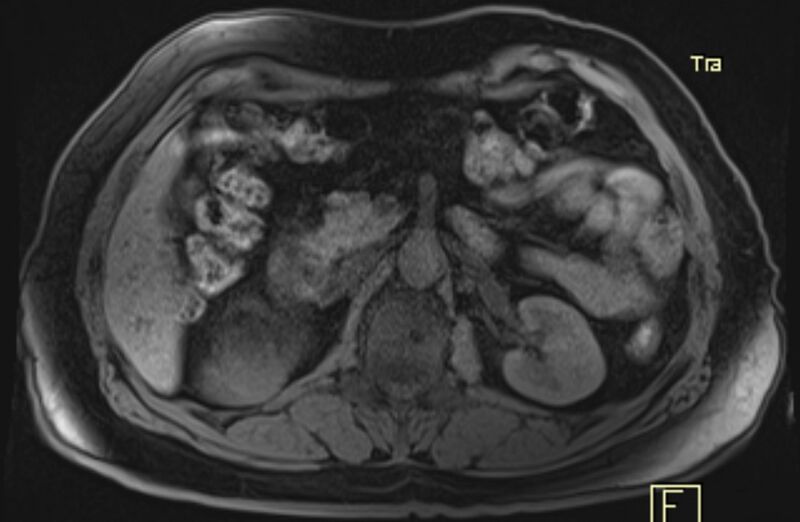
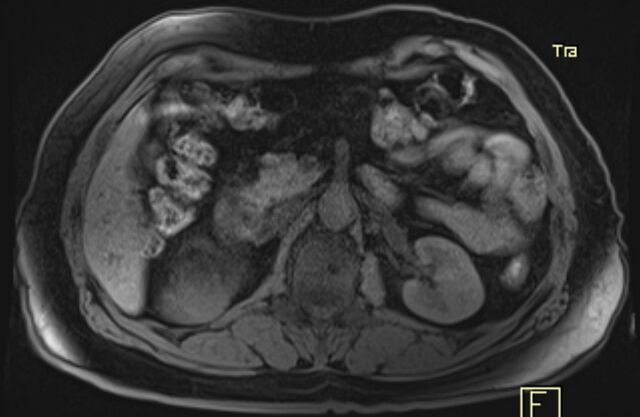
File:Adrenal myelolipoma (Radiopaedia 65240-74254 Axial T1 C+ fat sat 5).jpg
Jump to navigation
Jump to search

Size of this preview: 800 × 522 pixels. Other resolutions: 320 × 209 pixels | 640 × 417 pixels | 1,170 × 763 pixels.
Original file (1,170 × 763 pixels, file size: 259 KB, MIME type: image/jpeg)
Summary:
| Description |
|
| Date | Published: 4th Feb 2019 |
| Source | https://radiopaedia.org/cases/adrenal-myelolipoma-28 |
| Author | Mostafa El-Feky |
| Permission (Permission-reusing-text) |
http://creativecommons.org/licenses/by-nc-sa/3.0/ |
Licensing:
Attribution-NonCommercial-ShareAlike 3.0 Unported (CC BY-NC-SA 3.0)
File history
Click on a date/time to view the file as it appeared at that time.
| Date/Time | Thumbnail | Dimensions | User | Comment | |
|---|---|---|---|---|---|
| current | 07:55, 26 April 2021 | | 1,170 × 763 (259 KB) | Fæ (talk | contribs) | Radiopaedia project rID:65240 (batch #1360-65 D5) |
You cannot overwrite this file.
File usage
There are no pages that use this file.
.jpg&oldid=218318)