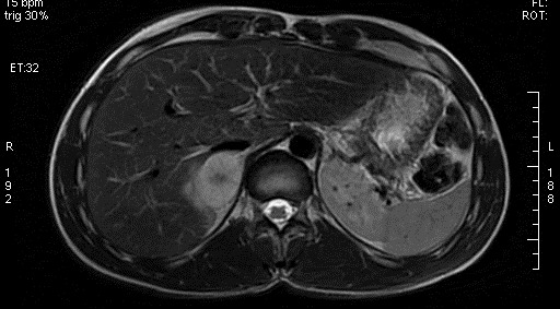
File:Adrenal pheochromocytoma (Radiopaedia 35133-36730 Axial T2 3).jpg
Jump to navigation
Jump to search
No higher resolution available.
Adrenal_pheochromocytoma_(Radiopaedia_35133-36730_Axial_T2_3).jpg (512 × 283 pixels, file size: 49 KB, MIME type: image/jpeg)
Summary:
| Description |
|
| Date | Published: 27th Nov 2015 |
| Source | https://radiopaedia.org/cases/adrenal-pheochromocytoma |
| Author | Essam G Ghonaim |
| Permission (Permission-reusing-text) |
http://creativecommons.org/licenses/by-nc-sa/3.0/ |
Licensing:
Attribution-NonCommercial-ShareAlike 3.0 Unported (CC BY-NC-SA 3.0)
File history
Click on a date/time to view the file as it appeared at that time.
| Date/Time | Thumbnail | Dimensions | User | Comment | |
|---|---|---|---|---|---|
| current | 17:30, 26 April 2021 | | 512 × 283 (49 KB) | Fæ (talk | contribs) | Radiopaedia project rID:35133 (batch #1384-3 A3) |
You cannot overwrite this file.
File usage
There are no pages that use this file.
.jpg&oldid=224459)