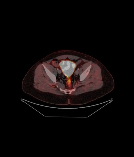
File:Adrenocortical carcinoma (Radiopaedia 80134-93440 ِAxial 234).jpg
Jump to navigation
Jump to search

Size of this preview: 514 × 600 pixels. Other resolutions: 206 × 240 pixels | 574 × 670 pixels.
Original file (574 × 670 pixels, file size: 60 KB, MIME type: image/jpeg)
Summary:
| Description |
|
| Date | Published: 27th Nov 2020 |
| Source | https://radiopaedia.org/cases/adrenocortical-carcinoma-4 |
| Author | Sherif Mohsen |
| Permission (Permission-reusing-text) |
http://creativecommons.org/licenses/by-nc-sa/3.0/ |
Licensing:
Attribution-NonCommercial-ShareAlike 3.0 Unported (CC BY-NC-SA 3.0)
File history
Click on a date/time to view the file as it appeared at that time.
| Date/Time | Thumbnail | Dimensions | User | Comment | |
|---|---|---|---|---|---|
| current | 20:23, 26 April 2021 |  | 574 × 670 (60 KB) | Fæ (talk | contribs) | Radiopaedia project rID:80134 (batch #1392-234 A234) |
You cannot overwrite this file.
File usage
The following page uses this file: