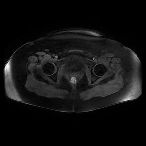
File:Adult granulosa cell tumor of the ovary (Radiopaedia 64991-73953 axial-T1 Fat sat post-contrast dynamic 15).jpg
Jump to navigation
Jump to search
Adult_granulosa_cell_tumor_of_the_ovary_(Radiopaedia_64991-73953_axial-T1_Fat_sat_post-contrast_dynamic_15).jpg (512 × 512 pixels, file size: 62 KB, MIME type: image/jpeg)
Summary:
| Description |
|
| Date | Published: 8th Jan 2019 |
| Source | https://radiopaedia.org/cases/adult-granulosa-cell-tumour-of-the-ovary |
| Author | Dr Ammar Haouimi |
| Permission (Permission-reusing-text) |
http://creativecommons.org/licenses/by-nc-sa/3.0/ |
Licensing:
Attribution-NonCommercial-ShareAlike 3.0 Unported (CC BY-NC-SA 3.0)
File history
Click on a date/time to view the file as it appeared at that time.
| Date/Time | Thumbnail | Dimensions | User | Comment | |
|---|---|---|---|---|---|
| current | 05:08, 27 April 2021 |  | 512 × 512 (62 KB) | Fæ (talk | contribs) | Radiopaedia project rID:64991 (batch #1415-134 F15) |
You cannot overwrite this file.
File usage
The following page uses this file:
