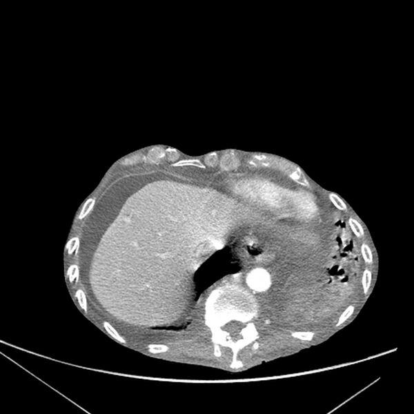
File:Advanced pulmonary tuberculosis (Radiopaedia 8599-9431 Axial liver window 17).jpg
Jump to navigation
Jump to search

Size of this preview: 600 × 600 pixels. Other resolutions: 240 × 240 pixels | 480 × 480 pixels | 768 × 768 pixels | 1,024 × 1,024 pixels.
Original file (1,024 × 1,024 pixels, file size: 144 KB, MIME type: image/jpeg)
Summary:
| Description |
|
| Date | Published: 15th Feb 2010 |
| Source | https://radiopaedia.org/cases/advanced-pulmonary-tuberculosis |
| Author | Natalie Yang |
| Permission (Permission-reusing-text) |
http://creativecommons.org/licenses/by-nc-sa/3.0/ |
Licensing:
Attribution-NonCommercial-ShareAlike 3.0 Unported (CC BY-NC-SA 3.0)
File history
Click on a date/time to view the file as it appeared at that time.
| Date/Time | Thumbnail | Dimensions | User | Comment | |
|---|---|---|---|---|---|
| current | 11:26, 27 April 2021 |  | 1,024 × 1,024 (144 KB) | Fæ (talk | contribs) | Radiopaedia project rID:8599 (batch #1442-17 A17) |
You cannot overwrite this file.
File usage
There are no pages that use this file.