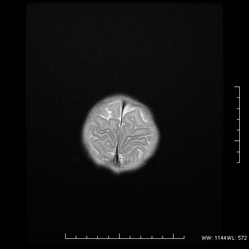
File:Agenesis of the corpus callosum (Radiopaedia 16190-16010 Axial T2 16).png
Jump to navigation
Jump to search
Agenesis_of_the_corpus_callosum_(Radiopaedia_16190-16010_Axial_T2_16).png (512 × 512 pixels, file size: 82 KB, MIME type: image/png)
Summary:
| Description |
|
| Date | Published: 28th Dec 2011 |
| Source | https://radiopaedia.org/cases/agenesis-of-the-corpus-callosum-1 |
| Author | Praveen Jha |
| Permission (Permission-reusing-text) |
http://creativecommons.org/licenses/by-nc-sa/3.0/ |
Licensing:
Attribution-NonCommercial-ShareAlike 3.0 Unported (CC BY-NC-SA 3.0)
File history
Click on a date/time to view the file as it appeared at that time.
| Date/Time | Thumbnail | Dimensions | User | Comment | |
|---|---|---|---|---|---|
| current | 15:56, 27 April 2021 |  | 512 × 512 (82 KB) | Fæ (talk | contribs) | Radiopaedia project rID:16190 (batch #1466-16 A16) |
You cannot overwrite this file.
File usage
There are no pages that use this file.
