File:Aggressive angiomyxoma (Radiopaedia 73343-84093 Sagittal T2 55).jpg

From NC Commons
Jump to navigation Jump to search

Aggressive_angiomyxoma_(Radiopaedia_73343-84093_Sagittal_T2_55).jpg(448 × 448 pixels, file size: 14 KB, MIME type: image/jpeg)

Summary:

Description
  • Radiopaedia case ID: 73343
  • Image ID: 51905069
  • Image stack position: 55/55
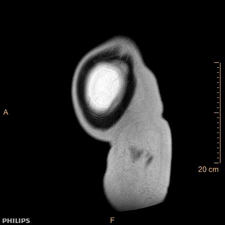
  • Plane projection: Sagittal
  • Aux modality: T2
  • Modality: MRI
  • System: Gynaecology
  • Findings: Unusual findings for a supposed ovarian cancer. ​Actually the main lesion originates from the subperitoneal space: it most likely comes from the perineum, breaks through the puborectal and levator ani muscles, extends into the right ischiorectal fossa and pushes to the left the rectum, uterus and bladder, without invading them. It then most likely extends in the retroperitoneal space up to the upper abdomen, behind the liver. The right colon is pushed away to the left, actually the entire digestive tract is pushed away as a whole. The lesion features mostly a liquid-like content, probably myxoid, (NOT ascites), T2 hyperintense, T1 hypointense with small T1FS hyperintense spots (blood or proteins), without fat.Interestingly there are long thin "filaments" enhancing after contrast injection. These are oriented vertically in a layered "onion-peel"-like fashion.
Date Published: 13th Jan 2020
Source https://radiopaedia.org/cases/aggressive-angiomyxoma
Author Fabien Ho
Permission
(Permission-reusing-text)
http://creativecommons.org/licenses/by-nc-sa/3.0/

Licensing:

Attribution-NonCommercial-ShareAlike 3.0 Unported (CC BY-NC-SA 3.0)

File history

Click on a date/time to view the file as it appeared at that time.

Date/TimeThumbnailDimensionsUserComment
current22:33, 27 April 2021Thumbnail for version as of 22:33, 27 April 2021448 × 448 (14 KB) (talk | contribs)Radiopaedia project rID:73343 (batch #1485-55 A55)