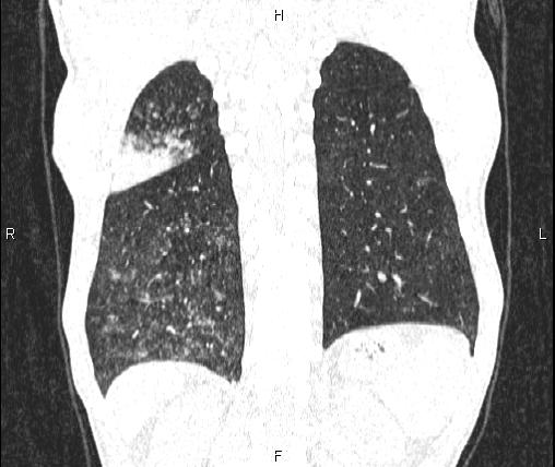
File:Air bronchogram in pneumonia (Radiopaedia 85719-101512 Coronal lung window 48).jpg
Jump to navigation
Jump to search
Air_bronchogram_in_pneumonia_(Radiopaedia_85719-101512_Coronal_lung_window_48).jpg (508 × 428 pixels, file size: 33 KB, MIME type: image/jpeg)
Summary:
| Description |
|
| Date | Published: 8th Jan 2021 |
| Source | https://radiopaedia.org/cases/air-bronchogram-in-pneumonia |
| Author | Mohammad Taghi Niknejad |
| Permission (Permission-reusing-text) |
http://creativecommons.org/licenses/by-nc-sa/3.0/ |
Licensing:
Attribution-NonCommercial-ShareAlike 3.0 Unported (CC BY-NC-SA 3.0)
File history
Click on a date/time to view the file as it appeared at that time.
| Date/Time | Thumbnail | Dimensions | User | Comment | |
|---|---|---|---|---|---|
| current | 18:44, 28 April 2021 |  | 508 × 428 (33 KB) | Fæ (talk | contribs) | Radiopaedia project rID:85719 (batch #1509-214 C48) |
You cannot overwrite this file.
File usage
The following page uses this file:
