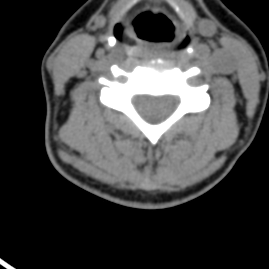
File:Alar ligament calcification (Radiopaedia 72589-83147 Axial liver window 26).jpg
Jump to navigation
Jump to search
Size of this preview: 600 × 600 pixels. Other resolutions: 240 × 240 pixels | 480 × 480 pixels | 874 × 874 pixels.
Original file (874 × 874 pixels, file size: 76 KB, MIME type: image/jpeg)
Summary:
| Description |
|
| Date | Published: 2nd Dec 2019 |
| Source | https://radiopaedia.org/cases/alar-ligament-calcification |
| Author | Antonio Rodrigues de Aguiar Neto |
| Permission (Permission-reusing-text) |
http://creativecommons.org/licenses/by-nc-sa/3.0/ |
Licensing:
Attribution-NonCommercial-ShareAlike 3.0 Unported (CC BY-NC-SA 3.0)
File history
Click on a date/time to view the file as it appeared at that time.
| Date/Time | Thumbnail | Dimensions | User | Comment | |
|---|---|---|---|---|---|
| current | 01:14, 29 April 2021 | | 874 × 874 (76 KB) | Fæ (talk | contribs) | Radiopaedia project rID:72589 (batch #1532-79 C26) |
You cannot overwrite this file.
File usage
There are no pages that use this file.
.jpg&oldid=245915)