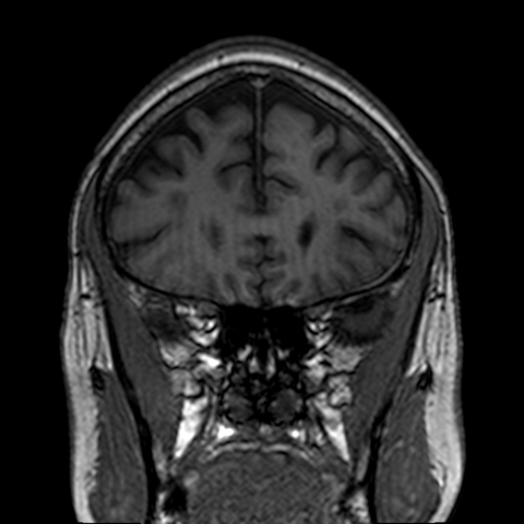
File:Alzheimeir's disease - probable (Radiopaedia 37261-39056 Coronal T1 17).png
Jump to navigation
Jump to search

Size of this preview: 600 × 600 pixels. Other resolutions: 240 × 240 pixels | 480 × 480 pixels | 768 × 768 pixels.
Original file (768 × 768 pixels, file size: 394 KB, MIME type: image/png)
Summary:
| Description |
|
| Date | Published: 14th Jun 2015 |
| Source | https://radiopaedia.org/cases/alzheimeirs-disease-probable |
| Author | Bruno Di Muzio |
| Permission (Permission-reusing-text) |
http://creativecommons.org/licenses/by-nc-sa/3.0/ |
Licensing:
Attribution-NonCommercial-ShareAlike 3.0 Unported (CC BY-NC-SA 3.0)
File history
Click on a date/time to view the file as it appeared at that time.
| Date/Time | Thumbnail | Dimensions | User | Comment | |
|---|---|---|---|---|---|
| current | 21:02, 29 April 2021 | | 768 × 768 (394 KB) | Fæ (talk | contribs) | Radiopaedia project rID:37261 (batch #1593-97 D17) |
You cannot overwrite this file.
File usage
The following page uses this file:
.png&oldid=252456)