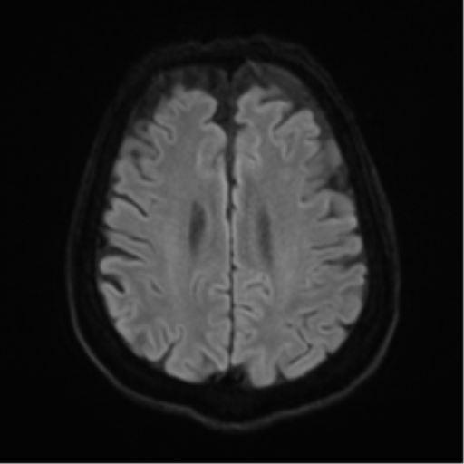
File:Alzheimer disease (Radiopaedia 57096-63994 Axial DWI 62).png
Jump to navigation
Jump to search
Alzheimer_disease_(Radiopaedia_57096-63994_Axial_DWI_62).png (512 × 512 pixels, file size: 51 KB, MIME type: image/png)
Summary:
| Description |
|
| Date | Published: 7th Dec 2017 |
| Source | https://radiopaedia.org/cases/alzheimer-disease-2 |
| Author | Bruno Di Muzio |
| Permission (Permission-reusing-text) |
http://creativecommons.org/licenses/by-nc-sa/3.0/ |
Licensing:
Attribution-NonCommercial-ShareAlike 3.0 Unported (CC BY-NC-SA 3.0)
File history
Click on a date/time to view the file as it appeared at that time.
| Date/Time | Thumbnail | Dimensions | User | Comment | |
|---|---|---|---|---|---|
| current | 22:48, 29 April 2021 |  | 512 × 512 (51 KB) | Fæ (talk | contribs) | Radiopaedia project rID:57096 (batch #1595-295 D62) |
You cannot overwrite this file.
File usage
The following page uses this file:
