File:Alzheimer disease and cerebral amyloid angiopathy (Radiopaedia 64234-73011 Coronal T1 33).png
Jump to navigation
Jump to search
No higher resolution available.
Alzheimer_disease_and_cerebral_amyloid_angiopathy_(Radiopaedia_64234-73011_Coronal_T1_33).png (512 × 512 pixels, file size: 224 KB, MIME type: image/png)
Summary:
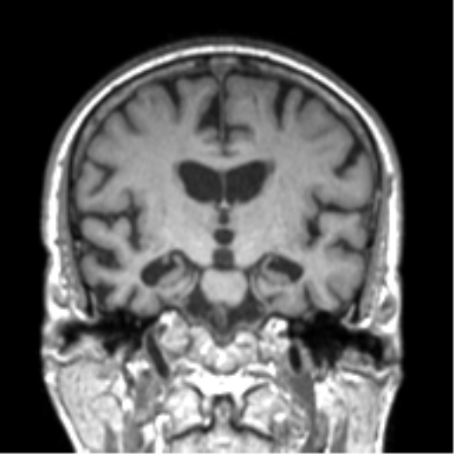
| Description |
|
| Date | Published: 11th Nov 2018 |
| Source | https://radiopaedia.org/cases/alzheimer-disease-and-cerebral-amyloid-angiopathy |
| Author | Frank Gaillard |
| Permission (Permission-reusing-text) |
http://creativecommons.org/licenses/by-nc-sa/3.0/ |
Licensing:
Attribution-NonCommercial-ShareAlike 3.0 Unported (CC BY-NC-SA 3.0)
File history
Click on a date/time to view the file as it appeared at that time.
| Date/Time | Thumbnail | Dimensions | User | Comment | |
|---|---|---|---|---|---|
| current | 00:24, 30 April 2021 | | 512 × 512 (224 KB) | Fæ (talk | contribs) | Radiopaedia project rID:64234 (batch #1598-115 C33) |
You cannot overwrite this file.
File usage
The following page uses this file:
.png&oldid=253736)