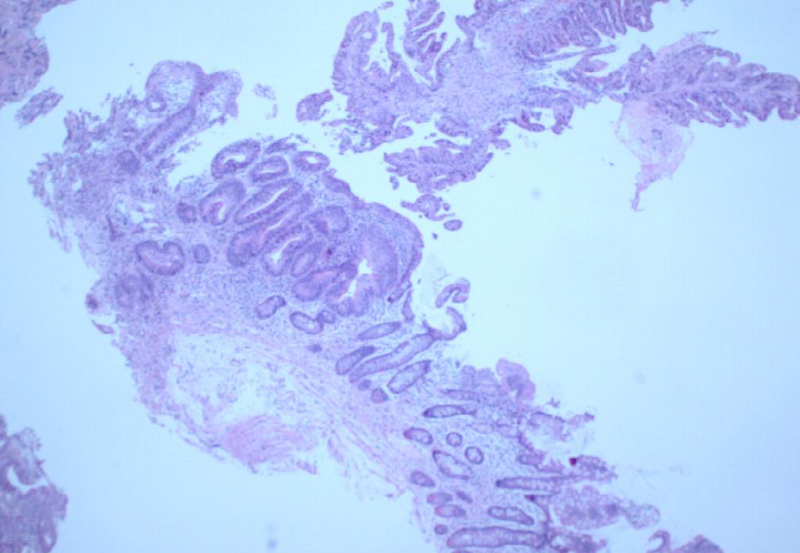
File:Ampullary adenocarcinoma (Radiopaedia 34013-35254 H&E 1).png
Jump to navigation
Jump to search

Size of this preview: 800 × 553 pixels. Other resolutions: 320 × 221 pixels | 640 × 442 pixels | 1,011 × 699 pixels.
Original file (1,011 × 699 pixels, file size: 830 KB, MIME type: image/png)
Summary:
| Description |
|
| Date | Published: 2nd Feb 2015 |
| Source | https://radiopaedia.org/cases/ampullary-adenocarcinoma |
| Author | Jan Frank Gerstenmaier |
| Permission (Permission-reusing-text) |
http://creativecommons.org/licenses/by-nc-sa/3.0/ |
Licensing:
Attribution-NonCommercial-ShareAlike 3.0 Unported (CC BY-NC-SA 3.0)
File history
Click on a date/time to view the file as it appeared at that time.
| Date/Time | Thumbnail | Dimensions | User | Comment | |
|---|---|---|---|---|---|
| current | 04:50, 1 May 2021 | | 1,011 × 699 (830 KB) | Fæ (talk | contribs) | Radiopaedia project rID:34013 (batch #1665-1 A1) |
You cannot overwrite this file.
File usage
There are no pages that use this file.
.png&oldid=263967)