File:Ampullary carcinoma (Radiopaedia 56396-63056 C 80).jpg

From NC Commons
Jump to navigation Jump to search

Ampullary_carcinoma_(Radiopaedia_56396-63056_C_80).jpg(512 × 512 pixels, file size: 43 KB, MIME type: image/jpeg)

Summary:

Description
  • Radiopaedia case ID: 56396
  • Image ID: 33494575
  • Image stack position: 80/111
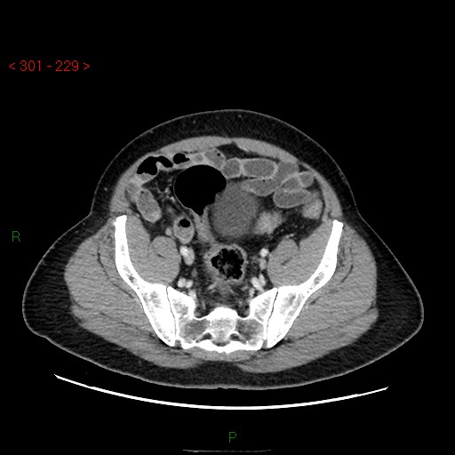
  • Plane projection: Axial
  • Aux modality: C+ portal venous phase
  • Modality: CT
  • System: Hepatobiliary
  • Findings: A tiny nodule was seen in the right middle lobe. A hypovascular mass of ampulla and associated marked dilatation of common bile duct and intrahepatic bile ducts. The pancreatic duct is prominent and not dilated. Normal fat planes preserved around major abdominal aortic branches. No sizable lymphadenopathy. The liver is average size and density with no definite suspicious focal lesions. The pancreatic head, neck, body and tail showing normal enhancement with no definite suspicious lesions seen Unremarkable CT features of the portal vein, spleen, adrenals, kidneys, urinary bladder, and visualized bowel loops. No ascites were seen. The visualized osseous structures showing small bony defect seen in the right iliac bone with an adjacent small bony projection which appearing of benign nature could be due to the previous operation for clinical history correlation with no definite suspicious bony lesions.
Date Published: 23rd Nov 2017
Source https://radiopaedia.org/cases/ampullary-carcinoma
Author Safwat Mohammad Almoghazy
Permission
(Permission-reusing-text)
http://creativecommons.org/licenses/by-nc-sa/3.0/

Licensing:

Attribution-NonCommercial-ShareAlike 3.0 Unported (CC BY-NC-SA 3.0)

File history

Click on a date/time to view the file as it appeared at that time.

Date/TimeThumbnailDimensionsUserComment
current09:29, 1 May 2021Thumbnail for version as of 09:29, 1 May 2021512 × 512 (43 KB) (talk | contribs)Radiopaedia project rID:56396 (batch #1670-164 C80)

The following page uses this file:

Metadata