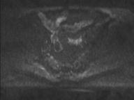
File:Anal adenocarcinoma - tumor regression grade 1 (Radiopaedia 31358-32100 Axial DWI 56).jpg

From NC Commons
Jump to navigation Jump to search

Anal_adenocarcinoma_-_tumor_regression_grade_1_(Radiopaedia_31358-32100_Axial_DWI_56).jpg(192 × 144 pixels, file size: 3 KB, MIME type: image/jpeg)

Summary:

Description
  • Radiopaedia case ID: 31358
  • Image ID: 8575944
  • Image stack position: 56/90
  • Plane projection: Axial
  • Aux modality: DWI
  • Study description: Post chemoradiotherapy
  • Study findings: Comparison is made with the previous examination. At the site of the previously identified anal lesion, no clearly viable residual tumor is identified. At the point of previously identified contact with the internal sphincter at the 3 o'clock position, there is a 17.7 x 2.3 x 11.6 mm lesion with no definite high T2 signal identified to suggest active tumor identified. Previously the lesion measured 25.5 x 5.9 x 20 mm. The rectum and anal sphincters appears normal. Since previous, the endometrial thickness has normalized. 1.5 centimeter posterior loops of uterine leiomyoma is again noted. Conclusion: Apparent excellent tumor response with significant reduction in size and no definite residual tumor signal (mrTRG 1).
  • Modality: MRI
  • System: Gastrointestinal
  • Findings: MR Anal Cancer Staging Multiplanar noncontrast small field of view MR imaging of the pelvis has been obtained. An anal polypoid lesion identified, extending down from the left anal mucosa. This measures 3.8 cm in length. Within the distal half of the anal canal, this lesion involves the mucosa at the 1-2 o'clock position, just touching the internal sphincter. The external sphincter is normal and the intersphincteric plane is preserved. The mesorectum and rectum appear normal. There are no abnormal inguinal lymph nodes. Visible bones are unremarkable. A 1.5 centimeter uterine leiomyoma is shown. In addition, there is increased endometrial thickness at 1.4 centimeters with small cystic spaces within, is seen in previous CT. Conclusion: Low anal canal lesion from the left side, probably just involving internal sphincter at the 1-2 o'clock position superficially at the level of the anal verge (T2). No external sphincter or intersphincteric fat plane involvement identified. No inguinal lymphadenopathy. Endometrial abnormality suspicious for malignancy, requires further evaluation.
Date Published: 16th Oct 2014
Source https://radiopaedia.org/cases/anal-adenocarcinoma-tumour-regression-grade-1
Author Jan Frank Gerstenmaier
Permission
(Permission-reusing-text)
http://creativecommons.org/licenses/by-nc-sa/3.0/

Licensing:

Attribution-NonCommercial-ShareAlike 3.0 Unported (CC BY-NC-SA 3.0)

File history

Click on a date/time to view the file as it appeared at that time.

Date/TimeThumbnailDimensionsUserComment
current03:16, 2 May 2021Thumbnail for version as of 03:16, 2 May 2021192 × 144 (3 KB) (talk | contribs)Radiopaedia project rID:31358 (batch #1705-161 E56)