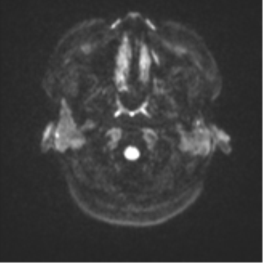
File:Anaplastic astroblastoma (Radiopaedia 55666-62194 Axial DWI 30).png
Jump to navigation
Jump to search
No higher resolution available.
Anaplastic_astroblastoma_(Radiopaedia_55666-62194_Axial_DWI_30).png (512 × 512 pixels, file size: 58 KB, MIME type: image/png)
Summary:
| Description |
|
| Date | Published: 25th Sep 2017 |
| Source | https://radiopaedia.org/cases/anaplastic-astroblastoma |
| Author | Ernest Lekgabe |
| Permission (Permission-reusing-text) |
http://creativecommons.org/licenses/by-nc-sa/3.0/ |
Licensing:
Attribution-NonCommercial-ShareAlike 3.0 Unported (CC BY-NC-SA 3.0)
File history
Click on a date/time to view the file as it appeared at that time.
| Date/Time | Thumbnail | Dimensions | User | Comment | |
|---|---|---|---|---|---|
| current | 04:07, 2 May 2021 | | 512 × 512 (58 KB) | Fæ (talk | contribs) | Radiopaedia project rID:55666 (batch #1712-246 E30) |
You cannot overwrite this file.
File usage
The following page uses this file:
.png&oldid=271174)