File:Anaplastic astroblastoma (Radiopaedia 55666-62201 H&E 1).jpg

From NC Commons
Jump to navigation Jump to search

Original file(960 × 720 pixels, file size: 165 KB, MIME type: image/jpeg)

Summary:

Description
  • Radiopaedia case ID: 55666
  • Image ID: 32805437
  • Aux modality: H&E
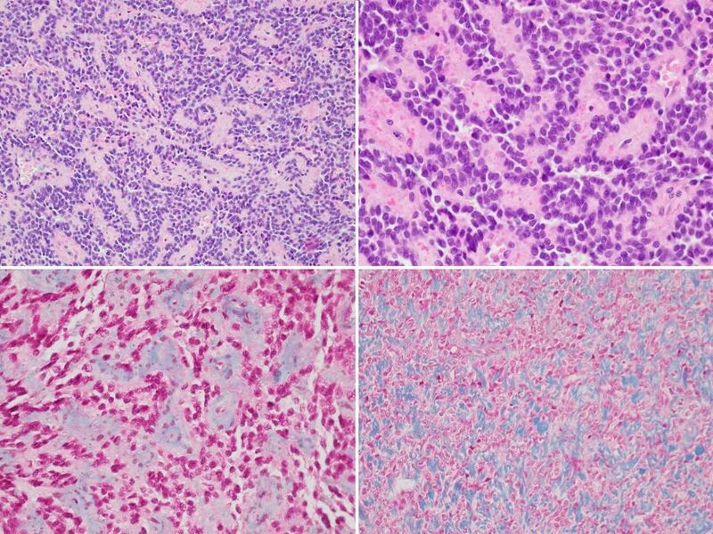
  • Study findings: MACROSCOPY The cut surface shows a homogeneous white rubbery appearance. No necrosis or hemorrhage are seen. MICROSCOPY: Sections show fragments of a well-circumscribed moderately hypercellular glioma. Focally tumor cells form papillae and perivascular rosettes with central hyalinised blood vessels. Tumor cells contain pale eosinophilic cytoplasm, oval nuclei with granular chromatin and inconspicuous nucleoli. Tumor cells demonstrate predominantly mild nuclear and cellular pleomorphism. Focally tumor cells show anaplastic features with enlarged, hyperchromatic nuclei and increased mitotic activity (up to 6/10hpf). No microvascular proliferation or pallisaded tumor necrosis are seen. Immunohistochemistry results show tumor cells stain: GFAP Positive Nestin Positive (high) NogoA Negative IDH-1 R132H Negative (not mutated) ATRX Positive (not mutated) EMA Positive (perinuclear dot-like) CD99 Positive NeuN Negative Synaptophysin Negative CyclinD1 Positive STAT6 Negative CD20 Negative Topoisomerase labeling index: Approximately 15-20%. Comment: The histological and immunohistochemical features are considered most consistent with Astroblastoma. The elevated mitotic count and high proliferation index both fulfill anaplastic criteria.
  • Modality: Pathology
  • System: Central Nervous System
  • Findings: 29 mm markedly T2 hypointense avidly enhancing lesion expanding cerebrocortex and involving the right precentral and inferior frontal gyrus laterally, and with marked diffusion restriction. The lesion is mostly solid with small cystic components peripherally. There is a focus of elevated cerebral blood volume (CBV) within the enhancing component. Extensive surrounding white matter T2 hyperintensity. Associated mass-effect causes partial effacement of the right lateral ventricle and midline shift of 5 mm to the left. No obstructive hydrocephalus. No transtentorial herniation.
Date Published: 25th Sep 2017
Source https://radiopaedia.org/cases/anaplastic-astroblastoma
Author Ernest Lekgabe
Permission
(Permission-reusing-text)
http://creativecommons.org/licenses/by-nc-sa/3.0/

Licensing:

Attribution-NonCommercial-ShareAlike 3.0 Unported (CC BY-NC-SA 3.0)

File history

Click on a date/time to view the file as it appeared at that time.

Date/TimeThumbnailDimensionsUserComment
current04:19, 2 May 2021Thumbnail for version as of 04:19, 2 May 2021960 × 720 (165 KB) (talk | contribs)Radiopaedia project rID:55666 (batch #1712-1 A1)

There are no pages that use this file.