File:Anaplastic astrocytoma (Radiopaedia 57768-64731 Axial DWI 13).png

From NC Commons
Jump to navigation Jump to search

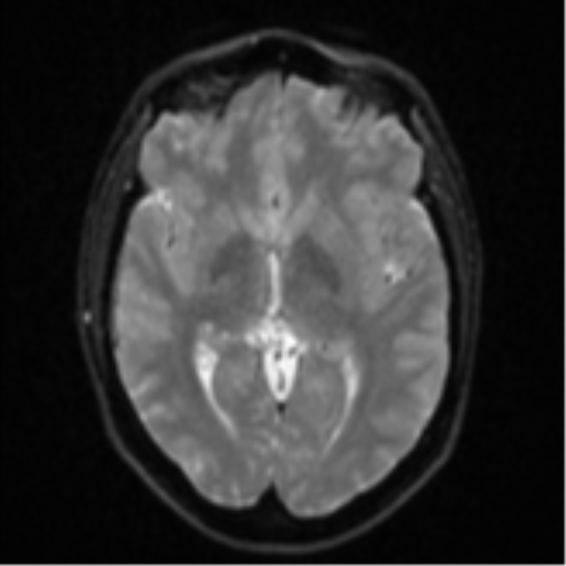
Anaplastic_astrocytoma_(Radiopaedia_57768-64731_Axial_DWI_13).png(512 × 512 pixels, file size: 69 KB, MIME type: image/png)

Summary:

Description
  • Radiopaedia case ID: 57768
  • Image ID: 35153407
  • Image stack position: 13/56
  • Plane projection: Axial
  • Aux modality: DWI
  • Study description: MRI Brain
  • Study findings: Intra-axial mass lesion centered on the left superior frontal gyrus measuring 2.7 x 2.6 cm with cortical expansion and diffuse surrounding white matter FLAIR/T2 hyperintense signal change within the left frontal lobe with the involvement of the body of the corpus callosum. The posterior component is more T2 hyperintense and FLAIR hypointense. No susceptibility. No definite restricted diffusion, but antero-medially there is a moderate size region of postcontrast enhancement (some areas gyriform) and with some increased CBV. There is resultant mass-effect with asymmetry of the left lateral ventricle and left-to-right shift of midline structures measuring 4 mm. Spectroscopy shows prominant choline peak with reduction of NAA, increased mI and mild lactate peak.
  • Modality: MRI
  • System: Central Nervous System
  • Findings: There is a large, ill-defined region of white matter hypoattenuation extending from the left frontal lobe with the involvement of the posterior left frontal cortical grey matter at the vertex. No definite enhancing mass or lesion is identified. No calcification. The associated mass effect causes moderate compression of the frontal horn of the left lateral ventricle, frontal sulcal effacement and 5 mm rightward shift of the anterior falx cerebri. No hydrocephalus and no transtentorial herniation. No acute intracranial hemorrhage or extra-axial collection. No suspicious osseous lesions. The imaged paranasal sinuses and mastoid air cells are clear. Conclusion: Ill-defined hypodense left frontal lesion with no definite enhancement is nonetheless suspicious for a brain neoplasm, most likely a primary tumor and unlikely to be a metastasis given the patient's age and pattern of involvement.
Date Published: 4th Feb 2018
Source https://radiopaedia.org/cases/anaplastic-astrocytoma-8
Author Bruno Di Muzio
Permission
(Permission-reusing-text)
http://creativecommons.org/licenses/by-nc-sa/3.0/

Licensing:

Attribution-NonCommercial-ShareAlike 3.0 Unported (CC BY-NC-SA 3.0)

File history

Click on a date/time to view the file as it appeared at that time.

Date/TimeThumbnailDimensionsUserComment
current06:13, 2 May 2021Thumbnail for version as of 06:13, 2 May 2021512 × 512 (69 KB) (talk | contribs)Radiopaedia project rID:57768 (batch #1714-188 G13)

Metadata