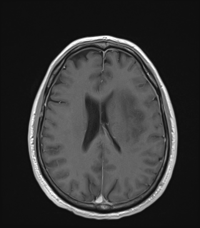
File:Anaplastic astrocytoma IDH wild-type (Radiopaedia 49984-55273 Axial T1 C+ 37).png
Jump to navigation
Jump to search
Anaplastic_astrocytoma_IDH_wild-type_(Radiopaedia_49984-55273_Axial_T1_C+_37).png (280 × 320 pixels, file size: 36 KB, MIME type: image/png)
Summary:
| Description |
|
| Date | Published: 14th Dec 2016 |
| Source | https://radiopaedia.org/cases/anaplastic-astrocytoma-idh-wild-type |
| Author | Bruno Di Muzio |
| Permission (Permission-reusing-text) |
http://creativecommons.org/licenses/by-nc-sa/3.0/ |
Licensing:
Attribution-NonCommercial-ShareAlike 3.0 Unported (CC BY-NC-SA 3.0)
File history
Click on a date/time to view the file as it appeared at that time.
| Date/Time | Thumbnail | Dimensions | User | Comment | |
|---|---|---|---|---|---|
| current | 09:43, 2 May 2021 |  | 280 × 320 (36 KB) | Fæ (talk | contribs) | Radiopaedia project rID:49984 (batch #1718-93 B37) |
You cannot overwrite this file.
File usage
The following page uses this file:
