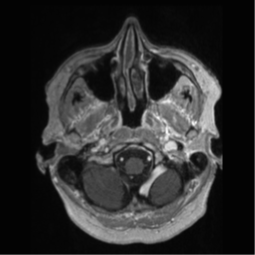
File:Anaplastic astrocytoma IDH wild-type (pseudoprogression) (Radiopaedia 42209-45277 Axial T1 C+ 9).png

From NC Commons
Jump to navigation Jump to search

Summary:

Description
  • Radiopaedia case ID: 42209
  • Image ID: 18725110
  • Image stack position: 9/133
  • Plane projection: Axial
  • Aux modality: T1 C+
  • Study description: 15 weeks post-surgery
  • Study findings: MRI obtained 15 weeks post surgery; Stupp protocol, finished 5 weeks earlier (first post treatment MRI). Right frontal craniotomy. The residual tumor in the right temporal frontal and parietal lobes has increased slightly in extent on T2-weighted images with slight increase in mass-effect with sulcal effacement and minor distortion of the right lateral ventricle. The tumor infiltrates the insula, posterior putamen, lateral thalmus, temporal lobes and extends superiorly into the right frontal and parietal lobes. A new area of peripheral contrast enhancement lies within the inferior frontal gyrus. Increased intensity of the regions of per contrast enhancement within the temporal lobe as well as around the region of necrosis are noted. However, the rCBV scan as shows less marked increase than on the prior study and the rADC shows less extensive regions of decreased ADC. MR spectroscopy shows areas of elevated lactate consistent with necrosis as well is persisting areas of elevated choline suggesting a residual tumor Conclusion: Although many of the imaging features suggest progressive disease, the reduction in rCBV as well as less extensive regions of decreased ADC suggest at least a partial response to therapy (pseudoprogression).
  • Modality: MRI
  • System: Central Nervous System
  • Findings: FLAIR hyperintensity in the right frontal parietal temporal region, with mild FLAIR hyperintensity extending to the ependymal surface of the right lateral ventricle posteriorly is noted, with areas of enhancement embedded within it. Although there is T2 shine through, there are also regions of true diffusion restriction, including at the areas of enhancement but also within the nonenhancing FLAIR hyperintensity. MRS demonstrates moderate choline elevation and small to moderate lactate peaks with reduced NAA. Elevation of cerebral blood volume (CBV) is prominent. Incidental left anterior temporal arachnoid cyst. Conclusion Features are those of a diffuse primary brain tumor. Increased cellularity and perfusion suggest a higher grade (III or IV).
Date Published: 15th Jan 2016
Source https://radiopaedia.org/cases/anaplastic-astrocytoma-idh-wild-type-pseudoprogression
Author Frank Gaillard
Permission
(Permission-reusing-text)
http://creativecommons.org/licenses/by-nc-sa/3.0/

Licensing:

Attribution-NonCommercial-ShareAlike 3.0 Unported (CC BY-NC-SA 3.0)

File history

Click on a date/time to view the file as it appeared at that time.

Date/TimeThumbnailDimensionsUserComment
current12:50, 2 May 2021Thumbnail for version as of 12:50, 2 May 2021512 × 512 (176 KB) (talk | contribs)Radiopaedia project rID:42209 (batch #1720-152 B9)

Metadata