File:Anaplastic astrocytoma IDH wild-type (pseudoprogression) (Radiopaedia 42209-45279 Axial DWI 21).png

From NC Commons
Jump to navigation Jump to search

Summary:

Description
  • Radiopaedia case ID: 42209
  • Image ID: 18725977
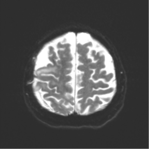
  • Image stack position: 21/54
  • Plane projection: Axial
  • Aux modality: DWI
  • Study description: 48 weeks post-surgery
  • Study findings: MRI obtained 11 months post op (3.5 months after last scan). Previous right parietal craniotomy and frontoparietal resection cavity again noted. The degree of FLAIR signal abnormality has reduced superiorly within the frontal and parietal lobes. Signal abnormality more inferiorly extending into the temporal lobe, insular and the right cerebral peduncle is similar to previous. The peripherally enhancing lobulated lesion is similar in extent to previous. The margins are less distinct, however no discrete new nodular enhancement is identified. There is persisting persisting reduced ADC values at the margins of the enhancement, progressed since previous with increased CBV within this region. New FLAIR signal abnormality involving the ipsilateral medial cerebral hemispheric cortex (precuneus and posterior cingulate) and contralateral (left) caudate head, anterior limb of internal capsule and the anterior putamen. This region does not demonstrate abnormal diffusion restriction or definite elevated CBV. Spectroscopy demonstrates elevated choline and reduced NAA within the FLAIR signal abnormality consistent with residual tumor. There is stable mass effect on the right cerebral hemisphere with stable ventricular size. Conclusion: While the extent of FLAIR signal abnormality within the superior frontal and parietal lobes has reduced, as has contrast enhancement, there is evidence of disease progression at the posteromedial margin of the resection cavity within the periventricular white matter as well as parietal and cingulate cortex. New mass like FLAIR abnormality involving left caudate head is also concerning for new tumor.
  • Modality: MRI
  • System: Central Nervous System
  • Findings: FLAIR hyperintensity in the right frontal parietal temporal region, with mild FLAIR hyperintensity extending to the ependymal surface of the right lateral ventricle posteriorly is noted, with areas of enhancement embedded within it. Although there is T2 shine through, there are also regions of true diffusion restriction, including at the areas of enhancement but also within the nonenhancing FLAIR hyperintensity. MRS demonstrates moderate choline elevation and small to moderate lactate peaks with reduced NAA. Elevation of cerebral blood volume (CBV) is prominent. Incidental left anterior temporal arachnoid cyst. Conclusion Features are those of a diffuse primary brain tumor. Increased cellularity and perfusion suggest a higher grade (III or IV).
Date Published: 15th Jan 2016
Source https://radiopaedia.org/cases/anaplastic-astrocytoma-idh-wild-type-pseudoprogression
Author Frank Gaillard
Permission
(Permission-reusing-text)
http://creativecommons.org/licenses/by-nc-sa/3.0/

Licensing:

Attribution-NonCommercial-ShareAlike 3.0 Unported (CC BY-NC-SA 3.0)

File history

Click on a date/time to view the file as it appeared at that time.

Date/TimeThumbnailDimensionsUserComment
current15:10, 2 May 2021Thumbnail for version as of 15:10, 2 May 2021512 × 512 (133 KB) (talk | contribs)Radiopaedia project rID:42209 (batch #1720-171 B21)

Metadata