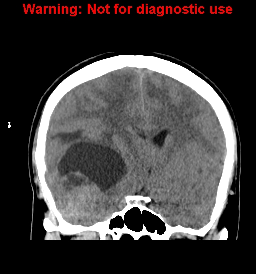
File:Anaplastic ganglioglioma (Radiopaedia 44921-48815 Coronal non-contrast 18).jpg
Jump to navigation
Jump to search
Anaplastic_ganglioglioma_(Radiopaedia_44921-48815_Coronal_non-contrast_18).jpg (512 × 549 pixels, file size: 108 KB, MIME type: image/jpeg)
Summary:
| Description |
|
| Date | Published: 10th May 2016 |
| Source | https://radiopaedia.org/cases/anaplastic-ganglioglioma |
| Author | Dr Amit Chakraborty |
| Permission (Permission-reusing-text) |
http://creativecommons.org/licenses/by-nc-sa/3.0/ |
Licensing:
Attribution-NonCommercial-ShareAlike 3.0 Unported (CC BY-NC-SA 3.0)
File history
Click on a date/time to view the file as it appeared at that time.
| Date/Time | Thumbnail | Dimensions | User | Comment | |
|---|---|---|---|---|---|
| current | 19:02, 2 May 2021 |  | 512 × 549 (108 KB) | Fæ (talk | contribs) | Radiopaedia project rID:44921 (batch #1730-79 C18) |
You cannot overwrite this file.
File usage
There are no pages that use this file.
