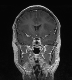
File:Anaplastic oligodendroglioma (Radiopaedia 37667-39563 Coronal T1 C+ 39).png
Jump to navigation
Jump to search
Anaplastic_oligodendroglioma_(Radiopaedia_37667-39563_Coronal_T1_C+_39).png (232 × 256 pixels, file size: 55 KB, MIME type: image/png)
Summary:
| Description |
|
| Date | Published: 18th Jun 2015 |
| Source | https://radiopaedia.org/cases/anaplastic-oligodendroglioma-2 |
| Author | RMH Neuropathology |
| Permission (Permission-reusing-text) |
http://creativecommons.org/licenses/by-nc-sa/3.0/ |
Licensing:
Attribution-NonCommercial-ShareAlike 3.0 Unported (CC BY-NC-SA 3.0)
File history
Click on a date/time to view the file as it appeared at that time.
| Date/Time | Thumbnail | Dimensions | User | Comment | |
|---|---|---|---|---|---|
| current | 05:40, 3 May 2021 |  | 232 × 256 (55 KB) | Fæ (talk | contribs) | Radiopaedia project rID:37667 (batch #1739-252 F39) |
You cannot overwrite this file.
File usage
The following page uses this file:
