File:Ancient neurilemmoma (Radiopaedia 33460-34509 A 18).png

From NC Commons
Jump to navigation Jump to search

Ancient_neurilemmoma_(Radiopaedia_33460-34509_A_18).png(512 × 512 pixels, file size: 130 KB, MIME type: image/png)

Summary:

Description
  • Radiopaedia case ID: 33460
  • Image ID: 10346195
  • Image stack position: 18/57
  • Plane projection: Axial
  • Aux modality: C+ arterial phase
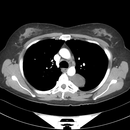
  • Study findings: There is a 37 x 65mm heterogeneous, partially calcified pleurally based mass in the left hemithorax abutting the proximal descending aorta. The mass partially erodes the posterior forth and fifth ribs and there is periosteal reaction on the anterosuperior edge of the forth rib. The mass is closely related to the intervertebral foramen of T4 and T5 however it is not widened. Subpleural ground glass opacity in the lingular segment of the left upper lobe is of uncertain significance. Mild dependent right sided atelectasis. Prominent mediastinal lymph nodes without lymphadenopathy by size criteria.
  • Modality: CT
  • System: Chest
  • Findings: Left paravertebral  based left upper zone mass located posteromedially with erosion of the inferior aspect of the posterior 4th rib. Lungs and pleural spaces are otherwise clear. Elevated left hemidiaphragm. Cardiomediastinal contour is within normal limits.
Date Published: 15th Jan 2015
Source https://radiopaedia.org/cases/ancient-neurilemmoma
Author Oliver Hennessy
Permission
(Permission-reusing-text)
http://creativecommons.org/licenses/by-nc-sa/3.0/

Licensing:

Attribution-NonCommercial-ShareAlike 3.0 Unported (CC BY-NC-SA 3.0)

File history

Click on a date/time to view the file as it appeared at that time.

Date/TimeThumbnailDimensionsUserComment
current16:44, 3 May 2021Thumbnail for version as of 16:44, 3 May 2021512 × 512 (130 KB) (talk | contribs)Radiopaedia project rID:33460 (batch #1782-18 A18)

The following page uses this file:

Metadata