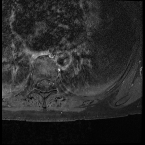
File:Ancient neurilemmoma (Radiopaedia 33460-34518 Axial T1 C+ fat sat 3).png

From NC Commons
Jump to navigation Jump to search

Ancient_neurilemmoma_(Radiopaedia_33460-34518_Axial_T1_C+_fat_sat_3).png(512 × 512 pixels, file size: 231 KB, MIME type: image/png)

Summary:

Description
  • Radiopaedia case ID: 33460
  • Image ID: 10349158
  • Image stack position: 3/29
  • Plane projection: Axial
  • Aux modality: T1 C+ fat sat
  • Study findings: A large, lobulated 4.4 X 7.9 (axial) X 4.8 (cc) cm T1 hypointense, heterogenously T2 hyperintense, enhancing, pleurally based left paraspinal mass posteromedially is demonstrated at the level of T4 and T5. There are areas of non-enhancement/cystic change within the mass, likely representing necrosis. Foci of susceptibility change within the mass are in keeping with calcification seen on CT. The mass abuts but doesn't involve of the left T4/5 intervertebral foramen. No overt rib destruction. There is minor pressure erosion (saucerisation) of the adjacent ribs and thoracic vertebra. This is not of pulmonary origin. A focal right paracentral T5/6 disc extrusion is present, which causes anterior indentation/effacement of the thecal sac. A smaller left paracentral left T6/7 disc protrusion also causes minor thecal sac indentation. No other significant spinal canal, lateral recess or foraminal stenosis.Marrow signal is normal. Vertebral alignment is unremarkable. No epidural mass or collection.Imaged lungs and other paraspinal structures are unremarkable, other than for dependent pulmonary atelectatic change. Conclusion Left T4/5 level pleurally-based mass as described, indeterminate. This is a long-standing lesion causing pressure remodeling of the adjacent vertebrae and ribs without invasion, remaining entirely extra-osseous. A solitary fibrous tumor is suspected. A chondral lesion or neural lesion are thought less likely.
  • Modality: MRI
  • System: Chest
  • Findings: Left paravertebral  based left upper zone mass located posteromedially with erosion of the inferior aspect of the posterior 4th rib. Lungs and pleural spaces are otherwise clear. Elevated left hemidiaphragm. Cardiomediastinal contour is within normal limits.
Date Published: 15th Jan 2015
Source https://radiopaedia.org/cases/ancient-neurilemmoma
Author Oliver Hennessy
Permission
(Permission-reusing-text)
http://creativecommons.org/licenses/by-nc-sa/3.0/

Licensing:

Attribution-NonCommercial-ShareAlike 3.0 Unported (CC BY-NC-SA 3.0)

File history

Click on a date/time to view the file as it appeared at that time.

Date/TimeThumbnailDimensionsUserComment
current17:10, 3 May 2021Thumbnail for version as of 17:10, 3 May 2021512 × 512 (231 KB) (talk | contribs)Radiopaedia project rID:33460 (batch #1782-83 D3)

There are no pages that use this file.
Metadata