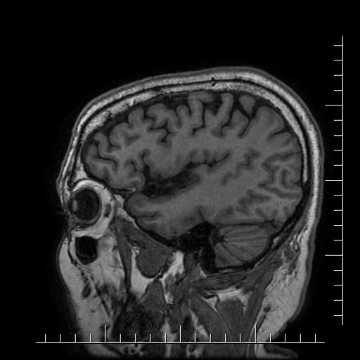
File:Aneurysm of posterior communicating artery (Radiopaedia 20188-20163 Sagittal T1 55).jpg
Jump to navigation
Jump to search
Aneurysm_of_posterior_communicating_artery_(Radiopaedia_20188-20163_Sagittal_T1_55).jpg (512 × 512 pixels, file size: 28 KB, MIME type: image/jpeg)
Summary:
| Description |
|
| Date | Published: 6th Nov 2012 |
| Source | https://radiopaedia.org/cases/aneurysm-of-posterior-communicating-artery |
| Author | Hani Makky Al Salam |
| Permission (Permission-reusing-text) |
http://creativecommons.org/licenses/by-nc-sa/3.0/ |
Licensing:
Attribution-NonCommercial-ShareAlike 3.0 Unported (CC BY-NC-SA 3.0)
File history
Click on a date/time to view the file as it appeared at that time.
| Date/Time | Thumbnail | Dimensions | User | Comment | |
|---|---|---|---|---|---|
| current | 16:36, 4 May 2021 |  | 512 × 512 (28 KB) | Fæ (talk | contribs) | Radiopaedia project rID:20188 (batch #1863-55 A55) |
You cannot overwrite this file.
File usage
The following page uses this file:
