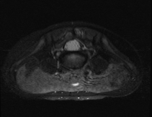
File:Aneurysmal bone cyst - sacrum (Radiopaedia 65190-74196 Axial T2 fat sat 2).jpg

From NC Commons
Jump to navigation Jump to search

Aneurysmal_bone_cyst_-_sacrum_(Radiopaedia_65190-74196_Axial_T2_fat_sat_2).jpg(512 × 394 pixels, file size: 37 KB, MIME type: image/jpeg)

Summary:

Description
  • Radiopaedia case ID: 65190
  • Image ID: 44991612
  • Image stack position: 2/20
  • Plane projection: Axial
  • Aux modality: T2 fat sat
  • Study findings: The MRI study better depicts the cystic nature of the sacral lesion. The lesion is bubbly-looking with multiple septations and fluid-fluid levels. It is arising from the posterior element of the sacral spine extending from S1 to S5 with mild extension to the body of S2 and S3 through the bilateral pedicles. There is significant narrowing at the distal sacral spinal canal, however, there is no compromise on the neural foramina. Minimal septal enhancement is seen in the post contrast images. The MRI features are highly suggestive of aneurysmal bone cyst.
  • Modality: MRI
  • System: Paediatrics
  • Findings: There is lytic lesion in the sacrum causing asymmetry of the sacral foramina. An overlapping fecal filled bowel loops degrade the image quality. The differential diagnosis of a midline sacral lesion in pediatric age group would include sacrococcygeal teratoma, chordoma, Ewing sarcoma or aneurysmal bone cyst. Further assessment with cross sectional imaging is needed to narrow the list.
Date Published: 28th Dec 2018
Source https://radiopaedia.org/cases/aneurysmal-bone-cyst-sacrum-1
Author Yasser Asiri
Permission
(Permission-reusing-text)
http://creativecommons.org/licenses/by-nc-sa/3.0/

Licensing:

Attribution-NonCommercial-ShareAlike 3.0 Unported (CC BY-NC-SA 3.0)

File history

Click on a date/time to view the file as it appeared at that time.

Date/TimeThumbnailDimensionsUserComment
current10:02, 4 May 2021Thumbnail for version as of 10:02, 4 May 2021512 × 394 (37 KB) (talk | contribs)Radiopaedia project rID:65190 (batch #1846-82 D2)

There are no pages that use this file.