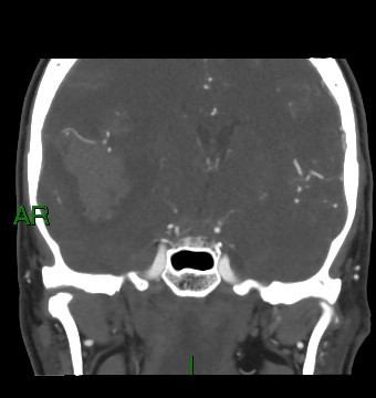
File:Aneurysmal subarachnoid hemorrhage with intra-axial extension (Radiopaedia 84371-99732 C 55).jpg

From NC Commons
Jump to navigation Jump to search

Aneurysmal_subarachnoid_hemorrhage_with_intra-axial_extension_(Radiopaedia_84371-99732_C_55).jpg(340 × 360 pixels, file size: 22 KB, MIME type: image/jpeg)

Summary:

Description
  • Radiopaedia case ID: 84371
  • Image ID: 53929023
  • Image stack position: 55/95
  • Plane projection: Coronal
  • Aux modality: C+ arterial phase
  • Study description: CT intracranial angiogram
  • Study findings: 6 mm aneurysm originating from the junction between the first and second segments of right middle cerebral artery. No other arterial aneurysm demonstrated. The pre-communicating segments of the posterior cerebral arteries bilaterally are relatively slender in caliber, with more normal caliber posterior communicating arteries and post-communicating segments of the posterior cerebral arteries.The previously demonstrated right frontal temporal hematoma is of similar size. Subarachnoid blood as before. Midline shift to the left and prominence of the temporal horns of the lateral ventricles with partial effacement of right lateral ventricle, as before.Opinion: 6 mm right middle cerebral artery aneurysm at the junction between the first and second segments.
  • Modality: CT
  • System: Central Nervous System
  • Findings: Right intra-axial hematoma within the right frontal-temporal region measuring 62 mm in maximum dimension. Peri hematoma edema.Subarachnoid blood in the basal cisterns, extending into the Sylvian fissures bilaterally and along the right frontal lobe sulci. Further subarachnoid blood in the pre-pontine cistern. No frank intraventricular blood. Near complete effacement of right lateral ventricle. Approximately 7 mm midline shift to the left. Prominence of the temporal horns particularly in the left lateral ventricle, is suggestive of a degree of early hydrocephalus. Likely right uncal herniation. No cerebellar tonsillar descent. No acute infarcts.Opinion: Extensive subarachnoid blood, and large right frontal temporal intra-axial hematoma. Given the extent of subarachnoid blood, the intra-axial hemorrhage more likely represents parenchymal extension of an aneurysmal subarachnoid hemorrhage rather than a primarily intra-axial hemorrhage with secondary subarachnoid extension - further evaluation with CT intracranial angiogram is advised.Likely early hydrocephalus and midline shift.
Date Published: 24th Nov 2020
Source https://radiopaedia.org/cases/aneurysmal-subarachnoid-haemorrhage-with-intra-axial-extension
Author Matthew Tse
Permission
(Permission-reusing-text)
http://creativecommons.org/licenses/by-nc-sa/3.0/

Licensing:

Attribution-NonCommercial-ShareAlike 3.0 Unported (CC BY-NC-SA 3.0)

File history

Click on a date/time to view the file as it appeared at that time.

Date/TimeThumbnailDimensionsUserComment
current16:05, 4 May 2021Thumbnail for version as of 16:05, 4 May 2021340 × 360 (22 KB) (talk | contribs)Radiopaedia project rID:84371 (batch #1860-184 C55)

Metadata