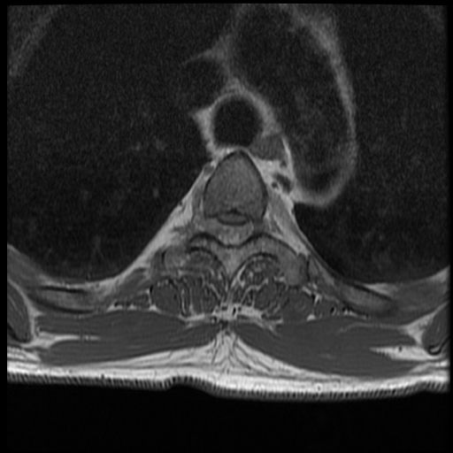
File:Angiolipoma - thoracic spine (Radiopaedia 28242-28479 Axial T1 23).jpg
Jump to navigation
Jump to search
Angiolipoma_-_thoracic_spine_(Radiopaedia_28242-28479_Axial_T1_23).jpg (512 × 512 pixels, file size: 107 KB, MIME type: image/jpeg)
Summary:
| Description |
|
| Date | Published: 19th Mar 2014 |
| Source | https://radiopaedia.org/cases/angiolipoma-thoracic-spine-1 |
| Author | Frank Gaillard |
| Permission (Permission-reusing-text) |
http://creativecommons.org/licenses/by-nc-sa/3.0/ |
Licensing:
Attribution-NonCommercial-ShareAlike 3.0 Unported (CC BY-NC-SA 3.0)
File history
Click on a date/time to view the file as it appeared at that time.
| Date/Time | Thumbnail | Dimensions | User | Comment | |
|---|---|---|---|---|---|
| current | 02:41, 5 May 2021 |  | 512 × 512 (107 KB) | Fæ (talk | contribs) | Radiopaedia project rID:28242 (batch #1880-51 C23) |
You cannot overwrite this file.
File usage
There are no pages that use this file.
