File:Ankle tendons- topographic anatomy (Radiopaedia 14238).jpg
Jump to navigation
Jump to search
Size of this preview: 800 × 600 pixels. Other resolutions: 320 × 240 pixels | 640 × 480 pixels | 1,024 × 768 pixels.
Original file (1,024 × 768 pixels, file size: 135 KB, MIME type: image/jpeg)
Summary:
- Radiopaedia case ID: 14238
- Image ID: 1191958
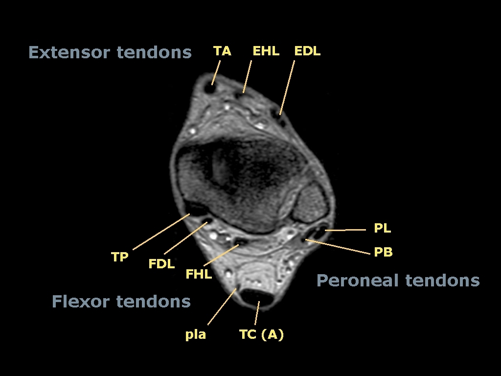
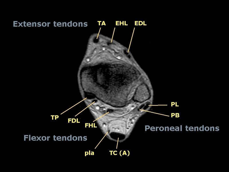
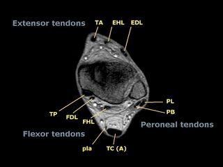
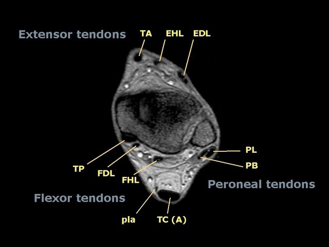
- Study findings: Transverse MRI section through the left ankle at the malleolar level showing the normal positions of the extrinsic tendons of the foot.
- Modality: MRI
- System: Musculoskeletal
- Findings: Transverse MRI section through the left ankle at the malleolar level showing the normal positions of the extrinsic tendons of the foot.
TA= tibialis anterior EHL= extensor hallucis longus EDL= extensor digitorum longus PL= peroneus longus PB= peroneus brevis TP= tibialis posterior FDL= flexor digitorum longus FHL= flexor hallucis longus TC(A)= tendo calcanei (Achilles) pla= plantaris tendon
- Published: 9th Jul 2011
- Source: https://radiopaedia.org/cases/ankle-tendons-topographic-anatomy
- Author: Roberto Schubert
- Permission: http://creativecommons.org/licenses/by-nc-sa/3.0/
Licensing:
Attribution-NonCommercial-ShareAlike 3.0 Unported (CC BY-NC-SA 3.0)
File history
Click on a date/time to view the file as it appeared at that time.
| Date/Time | Thumbnail | Dimensions | User | Comment | |
|---|---|---|---|---|---|
| current | 21:11, 18 March 2021 | | 1,024 × 768 (135 KB) | Fæ (talk | contribs) | Radiopaedia project rID:14238 (batch #1927) |
You cannot overwrite this file.
File usage
The following file is a duplicate of this file (more details):
.jpg)
.jpg)
The following page uses this file:
.jpg&oldid=8859876)