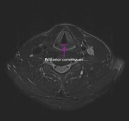
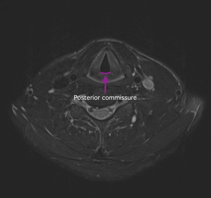
File:Anterior and posterior commissures of the larynx (diagrams) (Radiopaedia 61209-69116 B 1).jpg
Jump to navigation
Jump to search

Size of this preview: 639 × 600 pixels. Other resolutions: 256 × 240 pixels | 512 × 480 pixels | 818 × 768 pixels | 1,137 × 1,067 pixels.
Original file (1,137 × 1,067 pixels, file size: 301 KB, MIME type: image/jpeg)
Summary:
| Description |
|
| Date | Published: 23rd Jun 2018 |
| Source | https://radiopaedia.org/cases/anterior-and-posterior-commissures-of-the-larynx-diagrams |
| Author | Maxime St-Amant |
| Permission (Permission-reusing-text) |
http://creativecommons.org/licenses/by-nc-sa/3.0/ |
Licensing:
Attribution-NonCommercial-ShareAlike 3.0 Unported (CC BY-NC-SA 3.0)
File history
Click on a date/time to view the file as it appeared at that time.
| Date/Time | Thumbnail | Dimensions | User | Comment | |
|---|---|---|---|---|---|
| current | 14:18, 7 May 2021 | | 1,137 × 1,067 (301 KB) | Fæ (talk | contribs) | Radiopaedia project rID:61209 (batch #2100-2 B1) |
You cannot overwrite this file.
File usage
There are no pages that use this file.
_(Radiopaedia_61209-69116_B_1).jpg&oldid=309821)